This is an automatically translated article.
This article was written by Specialist Doctor II Khong Tien Dat, Doctor of Radiology - Department of Diagnostic Imaging - Vinmec Ha Long International Hospital. The doctor has a lot of experience with more than 14 years working in the field of diagnostic imaging.The tilting cardiopulmonary x-ray technique is often supplemented with the straight-line cardiopulmonary radiography technique, which helps doctors have more accurate bases for diagnosing patients with heart and lung diseases.
1. What is a tilting cardiopulmonary x-ray?
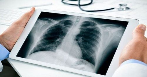
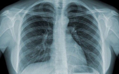
Tilt chest X-ray is an X-ray technique that uses an X-ray machine to record images of the heart, lungs, airways, and lymph nodes located inside the chest.There are two common types of chest X-ray: straight chest X-ray and inclined chest X-ray. In particular, the tilting technique plays a role in supplementing the results of the straight film, helping doctors have more bases to accurately diagnose patients with heart and lung diseases.
2. When to take a chest X-ray
Here are some signs that your lungs are weakening: A persistent cough; coughing up blood difficulty breathing; high fever ; chest pain after an injury such as a fracture or a pre-existing lung complication or signs of tuberculosis, lung cancer, or other heart or lung disease.When you notice the above signs, you need to see a doctor immediately and conduct a chest X-ray to assess the condition of many organs in the chest, whether there are any abnormalities or not?

3. Procedure for performing inclined chest X-ray
The procedure for performing cardiopulmonary radiographs is as follows:3.1 Preparations Specialist Doctor Electro-radiologist Equipment to prepare Specialized X-ray machine Film, film printer and storage system Patient side Patient Take off your shirt and remove metal objects on the body if it affects the technique, if necessary, the patient will put her hair neatly up. Test sheet The patient has an order to perform an X-ray from the doctor. Note: In case you are pregnant, you need to notify your doctor for a reasonable solution to avoid the influence of X-rays on the fetus in the abdomen.
3.2 Steps to conduct a tilting cardiopulmonary x-ray The following are the steps to perform a tilting cardiopulmonary x-ray:
The specialist receives an X-ray order from the patient. Conduct identification and compare the organ to be scanned with the clinical diagnosis. The patient enters the imaging room. The doctor will explain the imaging procedure to the patient Adjust the photograph, the shadow is 1.5m away from the photograph The doctor instructs the patient to stand on his side, his hands raised above his head, the chest to be photographed is close to the film Technician Adjust the center beam to the position at the lower border of the axillary D6 level. Stick the letter (F) to the digital sensor plate corresponding to the right of the patient. Enter the patient's name and age into the machine, select the program on the corresponding machine. corresponding to the organ to be scanned The technician adjusts (80kv, 8mAs corresponding to the patient) The doctor asks the patient to take a deep breath and hold his breath for a short time Close the door of the examination room and give the X-ray The patient leaves the room waiting for the results Inside the doctor and the radiologist adjust the contrast, check the balance of the images on the film. Print the film. Compare with the satisfactory film standards. 3.3 Interpretation of results On the X-ray film, the patient's name and age are recorded, the right and left stamps and the date of the film are taken. Entire lung: The apex of the lung and the flank angle of the diaphragm can be seen on both sides. The arm does not overlap the lung field. Full tilt: The sternum is completely tilted and the ribs are almost overlapping. Good deep inhalation: diaphragmatic arch below the anterior arch of the 6th rib. Hold breath well: contours of the heart and diaphragm are clearly seen. Good contrast: the light space behind the sternum is clearly visible, the light space behind the heart and the posterior diaphragmatic flank angle are clearly seen. The doctor analyzes the results displayed on the computer and prints the results. The doctor can provide professional advice if requested by the patient. 3.4 Complications and treatment

In some cases, it is necessary to repeat the technique if: the patient does not keep still during the scan, leading to blurred results, making it difficult for the doctor to read the results.
Please dial HOTLINE for more information or register for an appointment HERE. Download MyVinmec app to make appointments faster and to manage your bookings easily.
MOREDoes X-ray have any health effects? What are the benefits of magnetic resonance imaging (MRI)? 22 frequently asked questions about radiology


