This is an automatically translated article.
The article was professionally consulted by Specialist Doctor I Tran Cong Trinh - Radiologist - Radiology Department - Vinmec Central Park International General Hospital. The doctor has many years of experience in the field of diagnostic imaging.Arteriovenous malformations can occur anywhere in the body, resulting in malnutrition and dystrophy in some functional areas. Therefore, the method of vascular malformation of limbs through digital erasure angiography (DSA) is a technique being widely applied today to solve this problem.
1. What is limb malformation?
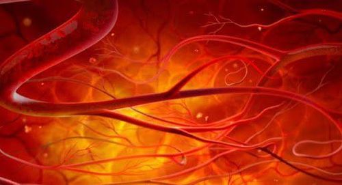
Arteriovenous malformation (AVM) is a congenital abnormality of blood vessels, formed during embryonic development. Arteriovenous malformations can occur anywhere in the body, from the central nervous system to peripheral soft tissue organizations.Vascular malformations include many direct connections between the arterial and venous systems without passing through the capillary system, thereby leading to a loss of metabolic capacity in the tissues surrounding the malformation, or Also called the phenomenon of blood robbery.
When the malformed arteriovenous and venous lesions progress according to the mature development of the body, it will lead to hypotrophy, tissue dystrophy causing manifestations: pain, deformity, ulceration - necrosis and bleeding .
Accessive endovascular intervention and embolization of limb malformations via arterial, venous or direct puncture will help restore circulation of the extremities, improve the nutritional status of downstream soft tissues.
2.Indications - Contraindications
Indication for embolization of limb vascular malformations Soft tissue arteriovenous malformations with complications: pain, skin dystrophy, ulceration, necrosis of surrounding soft tissue or downstream of the malformation Dynamic venous malformation The software has the effect of stealing blood downstream. Vascular embolization is contraindicated Patients with an allergy to iodine-containing contrast agents Patients with severe renal failure (grade IV) Patients with severe and uncontrolled coagulopathy (prothrombin index <60%, INR > 1.5 and platelet count < 50 g/l). Pregnant women.
Kỹ thuật chống chỉ định với phụ nữ có thai
3. The person who performs the DSA and node malformation technique
Specialist; Auxiliary physician; Electro-optical technician; Nursing; Doctor, anesthesiologist (if patient cannot cooperate).4. Means and drugs to prepare before taking DSA and embolization of limb malformations
Equipment and facilities for performing the procedure Digital angiography eraser (DSA); Dedicated electric pump; Film, film printer, image storage system; Lead shirt, apron to shield X-rays. Medicines to be prepared Local anesthetic; Pre-anesthesia and general anesthetic (if the patient has an indication for general anesthesia); Anticoagulants ; Drugs that neutralize anticoagulants; Water-soluble iodinated contrast agent; Antiseptic solution for skin and mucous membranes Prepare the patient The patient is thoroughly explained about the procedure and procedure to cooperate with the doctor and the technician; Patients need to fast, fast for 6 hours before or can drink no more than 50ml of water; After entering the intervention room, the patient will lie in the supine position, and the technicians will install a machine to monitor vital signs such as breathing rate, pulse, blood pressure, electrocardiogram, and arterial blood gases. In case the patient is uncooperative or aggressive, a sedative will be prescribed; Disinfect the skin surface with an antiseptic solution and cover with a sterile towel with holes.5. The procedure of imaging and digitizing the malformation of the digitized limb to erase the background
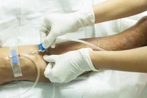
Người bệnh nằm ở tư thế ngửa trên bàn chụp DSA, kỹ thuật viên tiến hành đặt đường truyền
However, in exceptional cases where pre-anesthesia is required, such as: children under 5 years of age, who do not have a sense of cooperation with doctors, or patients who are too excited or scared... then it is necessary to conduct anesthesia. whole body during the procedure.
Intravascular catheterization Any malformation of any type requires a catheter to open the lumen to conduct angiography. If entering an artery or vein, use a 21G micro-needle set, then insert the tube into the lumen of the vessel 4-5Fr.
Angiography to assess lesions and embolization of limb vessel malformations Use catheters and microcatheters to access the arteriovenous malformations, then conduct non-selective and selective angiography to evaluate the type and hemodynamic status of the malformation.
Depending on the type of malformation, approach via artery, vein or direct puncture. Once the center of the malformation was accessed, angiography was performed to confirm the focal malformation and assess the hemodynamic status.
Carry out therapeutic vascular surgery by embolizing the malformed fovea with embolization material. Depending on the type and approach to the deformed drive, the appropriate material is selected. Angiography check after embolization, evaluate the adjacent and downstream branches, then close the path into the lumen, ending the intervention.
Focal deformity is successfully treated when the center of the fovea is occluded, the feeding vessel peduncles and draining vein cease to flow, and the adjacent and downstream vascular branches are preserved.
6. Complications when performing the procedure
Embolism downstream or adjacent vessels occurs with embolized materials that have the potential to migrate. Dissection or rupture of the vessel wall is rare but can occur in any stage of intervention. Cases of dissection upstream usually do not have serious consequences, but dissection downstream can cause diffuse dissection and embolism. Place a stent over the dissection site to correct. Pseudoaneurysmata at the site of vascular access is the most common complication, occurring in femoral artery access due to risk factors such as: weak vessel wall, atherosclerosis, loss of elasticity, patient mobility too early, the posterior femoral artery compression bandage is improperly intervened... Treated by embolization or suture surgery to restore the vessel wall. Complications cause vasospasm, need to be monitored for 10-15 minutes or can use selective vasodilators to manage; Accidents caused by broken/broken catheters or wires in the vascular lumen, then need to use special tools to remove through endovascular intervention or have to have surgery. Complications caused by contrast agents. Vinmec International General Hospital is a high-quality medical facility in Vietnam with a team of highly qualified medical professionals, well-trained, domestic and foreign, and experienced.A system of modern and advanced medical equipment, possessing many of the best machines in the world, helping to detect many difficult and dangerous diseases in a short time, supporting the diagnosis and treatment of doctors the most effective. The hospital space is designed according to 5-star hotel standards, giving patients comfort, friendliness and peace of mind.
Please dial HOTLINE for more information or register for an appointment HERE. Download MyVinmec app to make appointments faster and to manage your bookings easily.


