This is an automatically translated article.
Posted by Resident Doctor, Master Tran Duc Tuan - Department of Diagnostic Imaging - Vinmec Central Park International General HospitalThere are many ways to treat uterine leiomyoma, depending on the size of the tumor, the patient's clinical symptoms, etc. uterus. Each method has its own advantages and disadvantages.
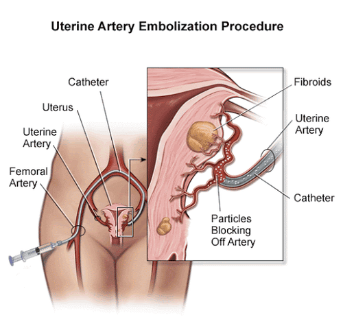
Uterine artery embolization in the treatment of cervical cancer has the following advantages: The procedure is relatively safe, the duration of the procedure and hospital stay is short, does not affect the patient's productivity in the future, and does not affect the patient's future. scars, as well as complications after surgery... Especially, the patient can get pregnant again. Uterine artery embolization is a method of inserting a catheter through the femoral artery into the internal iliac artery and into the uterine artery to inject substances that cause permanent embolism such as PVA plastic beads....
1. Indications and contraindications
1.1 Indications Uterine smooth muscle tumor less than 10cm in size, with clinical symptoms caused by the tumor such as: Abdominal pain, menorrhagia... If it is below the serosa, there is no stalk or attachment of the tumor. uterine muscle greater than or equal to 50% of the largest diameter of the tumor. Submucosal tumor < 5 cm in size. Uterine leiomyoma in women who want to preserve the uterus for childbirth or improve quality of life. Patients with UC with normal blood count, coagulation function, liver and kidney function, and normal vaginal cells. 1.2. Contraindications Uterine leiomyoma is too large, more than 10 cm in diameter. As with general contraindications to angiography: Presence of infection; liver failure, severe kidney failure; have hemophilia; diabetes mellitus; a history of allergy to preparations containing iodine; have a history of bronchial asthma ... Not pregnant, have appendicitis and suspected malignancy of the uterus and cervix.
2. Preparing to perform digital imaging techniques to erase the background and embolize uterine fibroids
2.1 Performer Interventional radiology specialist. Assistant doctor. Optical technician. Nursing. Doctor, anesthesiologist (if the patient cannot cooperate). 2.2 Media Film, film printer, image storage system Lead jacket, apron, X-ray shielding Digital angiography eraser (DSA) Dedicated electric pump. 2.3 Local anesthetics General anesthetics (if there is an indication for anesthesia). Skin and mucosal antiseptic solutions Anticoagulants Neutralizing anticoagulants Water soluble iodine contrast agents 2.4 Common consumables Syringe 1; 3; 5; 10 ml Syringe for electric pumps Distilled water or physiological saline Gloves, gown, cap, surgical mask Aseptic intervention kit: knife, scissors, tongs, 4 metal bowls, bean tray, Ins angiogram 4-5F Microcatheter 2-3F Micro-conductor 0.014-0.018 inch Guide Catheter 6F Y-connector set 2.6 Embolization material Bio-foam (hemostatic foam) Synthetic resin (PVA) Glue biology (Histoacryl, Onyx...) Metal spirals of all sizes (coils) 2.7 The patient is explained carefully about the procedure to coordinate with the doctor. Need to fast, drink before 6 hours. Can drink no more than 50 ml of water. In the intervention room: The patient lies on his back, fitted with a monitor to monitor breathing, pulse, blood pressure, electrocardiogram, SpO2. Disinfect the skin, then cover with a sterile, perforated cloth. The patient is too excited, can't lie still: Need to give sedation...
3. Technical process of digital imaging to erase the background and embolize the treatment of uterine fibroids
This technique is done in hospitals, patients only need to stay at the hospital after 1-2 days after the procedure. The patient was admitted to the hospital the day before the procedure and was thoroughly explained about the procedure for peace of mind. Before the procedure, the patient needs to be catheterized and defecated. Nursing the patient on the table, placing an intravenous line, placing an electrocardiogram and a vital function monitor, covering the large antiseptic genitals in the groin area on both sides. The doctor and the assistant wear lead coats, lead collars, wash hands, and wear gloves. Spread sterile gauze on the patient. Anesthetize the area of the common femoral artery 1 cm below the inguinal fold. Skin incision. Arterial puncture with a needle. Insert the catheter and catheter into the femoral artery. Insert the catheter into the uterine artery and take a scan, when it is satisfactory, pump PVA mixed with contrast material until the tumor is completely blocked, then stop. Take a re-check. Withdraw the catheter, insert it into the contralateral uterine artery and do the same as above. Remove catheter, arterial tube, compress the puncture site. The patient lies motionless for about 6-8 hours, after which the compression bandage can be removed. After embolization, antibiotics should be given to the patient to avoid infection. Monitoring after the technique:During the procedure: Monitor pulse, blood pressure After the procedure: monitor pulse, blood pressure, intelligence, pain level and give pain medication Super check negative after 3-6 -12 -24 months Magnetic resonance imaging can be done after 6 months. Evaluation of results: Complete occlusion of angiogenic tumor or partial depending on disease status, without loss of arterial branches 1/ 3 on the vagina as well as the vaginal artery.

4. Complications and treatment
Almost no serious complications occur May have the same complications as other angiograms: Bleeding, hematoma at the puncture site, but very rare Rare cases of necrosis of the cervical cyst with infection The patient may have pain lower abdomen after a few hours of procedure due to thrombosis, aseptic necrosis of the tumor Complications: There is no Vinmec International General Hospital with modern medical facilities, equipment and team Experts, doctors with many years of experience in medical examination and treatment, patients can completely rest assured to examine and treat Uterine leiomyoma at the Hospital.Please dial HOTLINE for more information or register for an appointment HERE. Download MyVinmec app to make appointments faster and to manage your bookings easily.
SEE MOREPrinciples of digital background angiography (DSA) Digital erasure of the splenic vein background - portal angiography Process of digitally erasing the background of the visceral system


