This is an automatically translated article.
Gill fissure disease is known to be one of the most common congenital diseases in children, accounting for about 30% of congenital tumors appearing in the neck region. Surgery to remove the gill fistula is currently the only method of treating the gill fistula.1.Surgery to remove cyst from gill fissure I
Cyst and fissure I is a rare malformation that is mainly caused by the normal overgrowth of gill slit I in the gill region during embryonic development. Until now, the origin of this malformation has not been determined, it is also possible that the failure of the closure of the gill fissure I leads to cysts and fistulas. Or it can also be explained by the inclusion of ectodermal traces, another theory is that it is the splitting of the external ear canal.
In fact, there are two different histological and anatomical types of cysts and oto- gill fistulas, depending on the relationship with the parotid gland, especially with the facial nerve.
The current method of treating fissure cysts is the surgical removal of slit fissures . The surgical removal of the fistula requires achieving the goal of removing all the fistula to avoid recurrence and complications.
What should be prepared before performing surgery to remove cyst from gill fissure I ?
Performer : Otolaryngologist trained in head and neck surgery.
Facilities:
Small scissors, ball, small trowel, small surgical forceps without anastomosis Bipolar electrode, electric knife Common surgical instruments. Can be retrofitted with a magnifying glass, microscope and wire monitor VII.

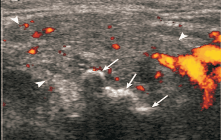
Người bệnh cần siêu âm tuyến mang tai trước khi thực hiện phẫu thuật lấy nang rò khe mang I
Patient: Need to clearly explain the risk of damage to the VII nerve, ask to conduct basic tests of a surgery. Perform a parotid ultrasound and if necessary, perform a fistula contrast-enhanced parotid tomography.
Technique of implementation: The skin incision is the same as in parotidectomy, extending downward in the cervical fold to the cyst or fistula. And this incision helps to preserve the branches of the VII cord and completely remove the cyst and fistula. The surgery was completed by making an opening into the outer ear canal, then removing a piece of the outer ear canal cartilage with the fistula.
2.Operating cysts and gill fissures II
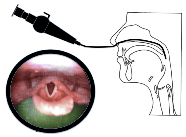
Cysts and fistulas of gill fissure II are caused by the existence of gill fissure II and cervical sinuses during embryonic development. However, during the development of the individual, these components will disappear. Before conducting cyst surgery and fistula II, what should be prepared? Performers: Otolaryngologists from specialty I and above and trained in head and neck surgery. Means:Local anesthetic (lidocaine + adrenaline 1/10,000). Software surgical kit. Patients: Conduct otolaryngoscopy examination as well as complete laboratory tests including complete blood count, baseline coagulation and liver and kidney function. Perform computed tomography scan of the neck in 2 coronal and axial positions. The anesthesiologist resuscitated the preoperative examination as well as explaining the procedure and possible complications.

Khám nội soi tai mũi họng cho bệnh nhân phẫu thuật nang và rò khe nang II
Technique:
Carry out subcutaneous anesthetic injection across the neck around the fistula with Medicaine (Octocaine) 1%. Make Skin incision through the skin attachment layer, peel the skin flap up and down according to the plane below the muscle attachment of the neck. Dissect the fistula around the fistula to find the fistula running in the carotid trough, the fistula that runs up to the level of the hyoid bone will usually go deep, that's why it is often necessary to make a second skin incision at the level of the great horn of the hyoid bone on the same side to get it. can easily remove the leak. For the fistula that runs deep into the tonsils at the superior border of the great horn of the hyoid bone, the fistula is removed to a deeper plane than the great horn of the hyoid bone, and the fistula is ligated. (Note: ligation of the fistula with non-absorbable sutures 2-0 or 3-0). The pathology of the cyst and the fistula of the gill slit is quite complicated, the surgical treatment is relatively difficult. Above are useful information about the surgery to remove the gill fissure cyst for readers to better understand the pathology of the gill slit.
Vinmec International General Hospital examines and treats common nasopharyngitis diseases, head and neck tumors, congenital malformations of the ear, nose and throat area with the most optimal internal and surgical methods for patients, both children and adults. Coming to Vinmec International General Hospital, patients will receive a direct, dedicated and professional examination from a team of qualified and experienced medical staff.
Any questions that need to be answered by a specialist doctor as well as customers wishing to be examined and treated at Vinmec International General Hospital, please register for an online examination on the Website for the best service.
Please dial HOTLINE for more information or register for an appointment HERE. Download MyVinmec app to make appointments faster and to manage your bookings easily.


