This is an automatically translated article.
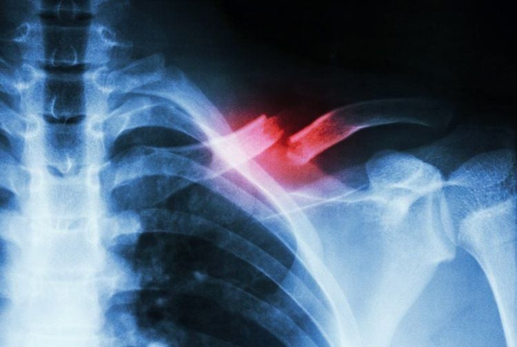
This article is professionally consulted by Master, Doctor Luu Thi Bich Ngoc - Radiologist - Department of Diagnostic Imaging and Nuclear Medicine - Vinmec Times City International Hospital.Collar fracture (clavicle) is the most common injury in the shoulder area, the rate of clavicle fracture accounts for 35-43% of shoulder fractures and 4% of whole body fractures. The cause of clavicle fracture is mainly due to falls, traffic accidents, 80% of the injury mechanism is due to indirect impacts such as when falling, hitting the shoulder, supporting the hand, the remaining 20% is caused by other impacts. direct impact, often causing open fracture.
1. What is a clavicle fracture?
A strong blow to the shoulder causes a broken collarbone or a fall in an outstretched arm position. Collar fractures can also occur from direct trauma to the collarbone from a traffic or other accident.Our body consists of 2 collarbones, which are bones located between the ribcage and shoulder blades, connecting the arms to the body.
The position of the collarbone lies on several important blood vessels and nerves. But when the collarbone is broken, these important structures are rarely injured, although when they break, the ends of the bones can be displaced.
The main cause of clavicle fracture is falls and traffic accidents. 80% of fracture injuries are caused by indirect trauma such as falling and hitting the shoulder, supporting the arm in the shoulder position. Direct trauma causes the remaining 20% and is usually an open fracture.
A clavicle injury is a fracture that heals easily. However, the belt will weaken if it is heavily deviated or there is no collarbone.
Collar fractures are usually closed fractures and do not cause any complications, or there are complications when the fracture is accompanied by some other damage, such as: damaged blood vessels, damaged nerves, membranes damaged lungs....or complicated clavicle fractures that cause the nerve bundle below the collarbone to be punctured, the top of the pleura is punctured, leading to pneumothorax. Those are the cases of clavicle fractures with complications.
It takes a certain amount of time for the bone to heal and recover. In conservative treatment, after 4-8 weeks the bone will have bone can. The patient will be pregnant during this time with conservative treatment.
For patients undergoing surgery to treat will be able to operate earlier thanks to the vehicle placed in the bone, without causing entanglement. Can bone is also affected by the process of bone dissection during surgery, so bone can form more slowly than conservative method.
Common symptoms caused by a collarbone fracture include:
Swelling, pain and bruising along the collarbone The pain increases and is felt when trying to move the shoulder or arm. “crack” sound The broken bone is deformed Falling forward or downward with the shoulder An infant with a collarbone fracture often cannot move the arm This is not a complete list of symptoms of a collarbone fracture cause. Therefore, if you have any unusual symptoms, you need to contact your doctor immediately.
You should see a doctor immediately if the following signs appear:
Numbness in your hand or a stinging sensation You feel very painful and cannot be treated with pain relievers Deformity in the shoulder and bone punctures the skin No can move the arm

2. Diagnosis of collarbone fracture
Collar fractures are usually diagnosed based on clinical presentation. However, doctors also often perform routine X-rays to accurately diagnose this condition. The patient is taken in an upright position, sometimes with the chest up or 45 degrees. However, with some fractures, additional imaging tests are required, such as computed tomography.The doctor will ask you more about the symptoms and the trauma situation in order to diagnose a collarbone fracture. By examining sensation and strength in your arms, fingers, and hands, your doctor will determine if nerve damage has occurred.
Doctors will recommend a collarbone X-ray for further diagnosis if a broken collarbone is suspected. The images obtained through X-rays will help the doctor determine if the collarbone is broken and determine the severity of the condition or whether other bones are affected.
3. Treatment of clavicle fracture
Because the clavicle has a thick periosteum, it is a type of bone that heals easily when broken because the clavicle has a thick periosteum and the upper position of the ribcage is an area of abundant blood supply, so the clavicle is easy to heal when broken. .There are 2 main methods when treating clavicle fractures, including: conservative treatment and surgical treatment.
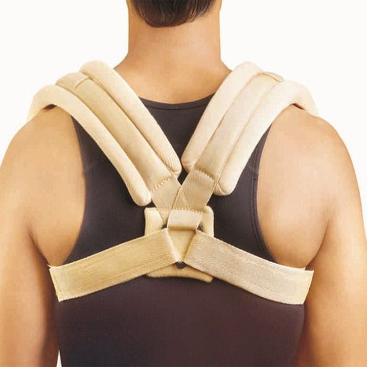
Conservative treatment The majority of clavicle fractures are treated conservatively with a belt. Previously, to adjust the patient's shoulder to tilt back, fix the collarbone, the patient would be cast in a cast. Today, patients can opt for gentle conservative treatment, meaning that using a
8 elastic when wearing a belt is more gentle for the patient.
For elderly patients, the doctor will also prescribe conservative treatment, bone fusion surgery will not be able to guarantee if the bones are already thinning. Or conservative treatment should be given to patients who do not accept great risks.
When the patient cannot accept the painful surgery or the surgical scar, and does not want to stay in the hospital...conservative treatment will be applied to these patients.
It is possible that the clavicle will not be able to heal and the bone can not grow when conservative treatment is not good. Or the broken bone protrudes, causing skin ulcers, punctures out due to the pressure preservation process.
The healing process of the conservative treatment does not achieve the absolute shape compared to the surgical method, leading to the appearance of deviations, ruffles, shortening of the shoulder, and protrusion of the clavicle. up, causing cosmetic loss to the patient.
Patients need to stay the same and visit the hospital regularly to overcome complications of conservative treatment. After about 1 week of conservative treatment, the patient should go to the hospital so that the doctor can check if the film is successful, there is a risk of bone piercing through the skin. The doctor will recommend surgery if the patient is likely to have complications.

If there is a third fracture with the risk of perforating the skin or pleura with closed fractures being treated conservatively, surgery will also be indicated.
Collar fractures in young people are often encouraged to have surgery to fuse the bones. The doctor will weigh the benefits and risks for the patient.
Compared with conservative treatment, patients will be better chiropractic when choosing surgery. However, compared with the conservative treatment method, the surgical method will leave the surgical scar and the surgical cost will be higher, and the patient will have to perform a second surgery to remove the medical instruments. outside. Therefore, patients need to consider between these two treatment methods.
In addition, due to the aesthetic needs of people, indications for surgery are increasingly being expanded. When conservative treatment has complications such as bone dislocation, due to the fact that the clavicle is located just below the skin, the healing process is not as expected, creating a raised tumor, causing aesthetic loss. Patients may require surgery to heal the bone if concerned about this.
Currently, X-ray is an imaging method that is performed routinely at Vinmec International General Hospital, including the technique of X-ray of the collarbone. The X-ray procedure at Vinmec is carried out methodically under the guidance of the medical team of the Department of Diagnostic Imaging. In addition, Vinmec is now equipped with the most modern X-ray machine, meeting international standards for true and clear images, helping doctors to accurately diagnose the disease and the stage of the disease, from It has an effective treatment direction, creating a sense of safety for the patient.
Any questions that need to be answered by a specialist doctor as well as customers wishing to be examined and treated at Vinmec International General Hospital, please register for an online examination on the Website for the best service.
Please dial HOTLINE for more information or register for an appointment HERE. Download MyVinmec app to make appointments faster and to manage your bookings easily.


