This is an automatically translated article.
The article is professionally consulted by Master, Doctor Ton Nu Tra My - Department of Diagnostic Imaging - Vinmec Central Park International General Hospital. The doctor has many years of experience in the field of diagnostic imaging.Ultrasound is a diagnostic imaging technique widely used in medicine. Today, ultrasound machines are becoming more and more complete and advanced, helping to improve patient safety and being an effective assistant for doctors in diagnosis and treatment. So what is the principle of imaging in medical ultrasound?
1. What is an ultrasound?
Ultrasound is one of the most widely used non-invasive imaging methods. The principle of imaging in ultrasound is to record normal or abnormal images of some organs in the body through the application of the mechanism of ultrasound waves and reconstruct the images by a modern computer system. .Through that, ultrasound both helps diagnose different diseases, and is valuable in monitoring and responding to patients' treatment. Ultrasound can also provide real-time images, so it can evaluate the structure and movement of organs in the body.
2. What is ultrasound used for?

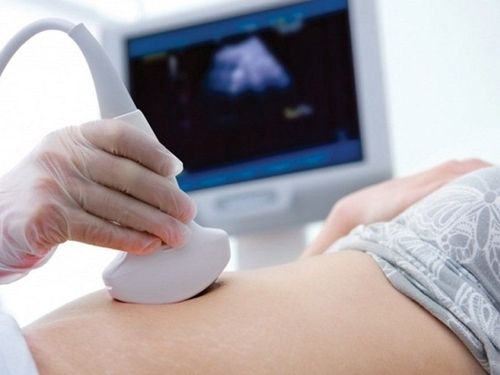
Siêu âm ổ bụng cho bệnh nhân
Survey of organs and organs such as general abdominal ultrasound, obstetrics and gynecology, cardiovascular, kidney, urology or prostate, thyroid ultrasound, mammography, musculoskeletal or testicular ultrasound... Besides diagnosis, ultrasound is also used to guide biopsies, pleural, peritoneal, or supportive treatment.
3. What is the working mechanism of ultrasound machine?
The main operating platform of the ultrasound machine is based on the principle of positioning by ultrasonic waves. When operating, through a special probe that both emits and receives the feedback of ultrasound waves, the doctor will place this probe close to the patient's skin to record images of the organs below.
Nguyên lý hoạt động cơ bản của máy siêu âm
Some of the other sound waves will continue to pass through, deeper into the body and meet other boundaries deeper, creating another acoustic feedback back to the transducer.
This special ultrasound transducer will receive different reflected sound waves, process and send the obtained information to a modern computer system for organ reconstruction. This is the principle of imaging in ultrasound.
The ultrasound machine works based on 2 parameters: the speed at which the sound waves travel in the tissue and the time it takes for the echo to return to the transducer. After being processed, the ultrasound machine will display the obtained images on the screen with different display modes.
Currently, most ultrasound machines will have the same operating principle. In order to provide clear images with no different dimensions, technicians have developed modern 3D and 4D ultrasound machines to make diagnosis and treatment easier.
4. Types of ultrasound image display
Type A (Amplitude Mode, A Mode): This display type also works on the basic principle of ultrasound. The transducer emits sound waves intermittently, when the sound waves pass through the tissues, different echoes are generated. From there, electrical signals are generated, which are processed by the computer and displayed on the screen as spikes that rise above the isoelectric line.Type B (Brightness Mode, B Mode): The principle is the same, but the resulting images will be displayed as grayscale in real time. The stronger the reflected signal, the brighter the resulting image. Tissues that give strong echoes are white, no echoes are black, different tissues give different echoes according to convention and are represented by gray scale colors.
TM mode (Time Motion Mode, TM Mode): This is essentially a combined B Mode ultrasound that displays organ movements in real time. TM Mode ultrasound is used to assess movement, measure size, elasticity...
5. Types of ultrasonic transducers

Hình ảnh một số loại đầu dò siêu âm
Then, when the reflected waves return, the piezoelectric ceramic pads vibrate and generate pulses, which are fed to the computer system for processing and image reconstruction. Based on different survey functions and frequencies, manufacturers will manufacture probes with suitable shapes and sizes:
Linear Array: This type of probe has high frequency, for high resolution images. Widely used in ultrasound of superficial areas such as skin, thyroid, mammary glands, blood vessels... Curved transducer (convex): This type has lower frequency, lower resolution image. Bonus use to evaluate deep organs such as abdominal cavity, fetal ultrasound or deep blood vessels... Echocardiogram transducer : Similar to a curved transducer and used exclusively in imaging evaluation of the heart.
6. Classification of ultrasound machines

Máy siêu âm GE E9
By shape: trolley ultrasound machine, portable (benchtop) ultrasound machine, hand-held machine. .. By technology: Black and white ultrasound machine, 3D/4D ultrasound machine, color ultrasound, Doppler ultrasound, ... According to the scope of application: Echocardiogram machine, general ultrasound machine, ultrasound machine Obstetrics/Gynecology... Vinmec International General Hospital system uses the most modern generations of color ultrasound machines to examine and treat patients. One of them is GE Healthcarecar's Logig E9 ultrasound machine with full options, HD resolution probes for clear images, accurate assessment of lesions. In addition, a team of experienced doctors and nurses will greatly assist in the diagnosis and early detection of abnormal signs of the body in order to provide timely treatment.
Please dial HOTLINE for more information or register for an appointment HERE. Download MyVinmec app to make appointments faster and to manage your bookings easily.