This is an automatically translated article.
The article is professionally consulted by Master, Doctor Ton Nu Tra My - Department of Diagnostic Imaging - Vinmec Central Park International General Hospital. The doctor has many years of experience in the field of diagnostic imaging.1. What is Tissue Elastography?
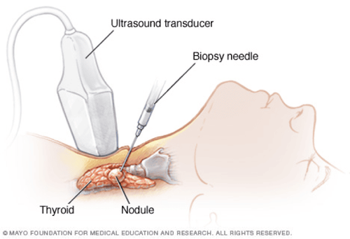
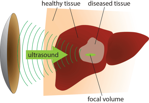
Currently, ultrasound diagnosis of thyroid and breast tumors is quite common, with the rate of thyroid cancer accounting for 2.1% of all cancers in Vietnam. Breast cancer ranks first in women. with an incidence rate of 19.7/100000 population/year. Early detection of cancer is essential for effective treatment of malignancies (if any).Tissue elastography is a modern and advanced method to help detect breast cancer, thyroid cancer, liver cirrhosis and soft tissue conditions early. Tissue elastography works on the principle of using pressure to displace tissue and track the movements caused in the tissue.
Tissue elastography makes the fastest and most effective diagnosis for tumors with high hardness, wide invasion and potential for malignancy. From the application of tissue elastography and TIRADS grading, the correct cancer diagnosis is 91.7%, the specificity is 98.3%.
2. Principle of tissue elastic ultrasound
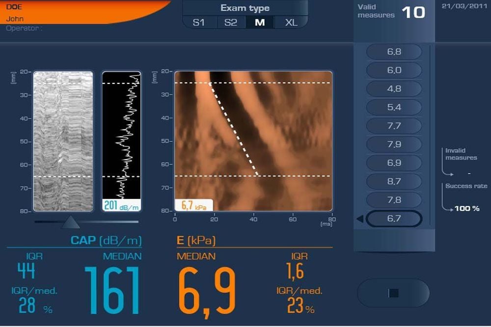
Changes in tissue elasticity or stiffness are both associated with pathological changes (benign or malignant). With the same force, soft tissue deforms more and hard tissue deforms less. Many types of soft tissue have the same echogenicity but have different stiffness.Elastography is performed by measuring the deformation of the lesion and is color coded into an elastic map (Elastogram). The extent of tissue displacement and change will be shown on the Elastogram, the color of the lesion will be superimposed on the B-mode image, the hard tissue will show up in black and the soft tissue will show up in white. The tumor is usually harder than the surrounding tissue, so it will appear as a black mass on a white background.
There are two main types of ultrasound waves:
Longitudinal waves: compressing the tissue, causing the tissue to deform, with a speed of ~1540m/s. Longitudinal waves are commonly found in solids and liquids. Transverse wave: generates shear wave with speed 0-10m/s. Transverse waves are found only in solids and liquid surfaces. As the stiffness of the tissue increases, the shear wave velocity increases. Thus, according to the principle of elastography, the distinction between benign and malignant lesions is based on:
Relative stiffness of the lesion on the Elastogram (Elasticity Score - ES) The ratio of the area of the lesion in the Elastogram to that of the Elastogram. with B-mode ultrasound image ( Area Ratio - AR ) The ratio of the deformation of the lesion compared to the surrounding tissue ( Strain Ratio - SR ).
3. Application of tissue elasticity ultrasound
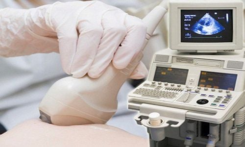
If B-mode ultrasound has high application in observing the anatomy of organs, Doppler ultrasound helps to evaluate blood flow, tissue elastography has advantages in diagnosing tissue stiffness.Ultrasound elastography is a modern method often indicated and applied in the following cases:
Ultrasound diagnosis of thyroid nodules. Ultrasound diagnosis of breast tumors, breast cancer. Ultrasound determines the degree of liver fibrosis in high-risk subjects such as: hepatitis B, fatty liver, metabolic diseases affecting the liver. Elastic ultrasound of spleen, kidney, pancreas, prostate...
Vinmec International General Hospital is one of the hospitals that not only ensures professional quality with a team of leading medical doctors, modern equipment and technology, but also stands out for its examination and consultation services. comprehensive and professional medical consultation and treatment; civilized, polite, safe and sterile medical examination and treatment space.
If you have a need for consultation and examination at Vinmec Hospitals under the nationwide health system, please book an appointment on the website for service.
Please dial HOTLINE for more information or register for an appointment HERE. Download MyVinmec app to make appointments faster and to manage your bookings easily.


