This is an automatically translated article.
The stomach is not contrasted with X-rays, so when taking X-rays of the stomach, the patient must take Barium (also known as Baryt) contrast agent. X-ray imaging of the stomach helps identify physical damage and disorders of the stomach, as well as signs of compression from the outside.
The article is professionally consulted by Master, Doctor Le Anh Viet - Radiologist - Department of Diagnostic Imaging and Nuclear Medicine - Vinmec Times City International General Hospital.
1. Normal stomach image on X-ray film
1.1. Location
The stomach is located just below the left diaphragm, consisting of 2 parts:
The vertical part: Almost parallel to the left side of the spine Transverse part: Crossing the spine, obliquely to the right. The stomach of a fat person is usually located high and runs horizontally above the navel, circulates quickly, and the two curves increase. The stomach of thin people and women is often low, circulation is slow and peristalsis is reduced.
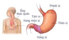
Vị trí dạ dày của người béo và người gầy có sự khác biệt
1.2. Shape
The air bag is located close to the lower border of the left diaphragm, on a standing gastric x-ray, air and fluid levels are seen. In some cases, the stomach has a waterfall shape, on the vertical film, two levels of gas and fluid will be seen in the gastric air sac. The cause of the waterfall stomach can be congenital, sometimes caused by an ulcer in the cardia.
1.3. Two curved banks
Greater curvature: Left border of stomach, from gastric aneurysm to pyloric canal. The mucosa at this site is often very rough, easily confused with pathological lesions. Small curvature: Right border of the stomach, running from the cardia to the pyloric canal. In which, the pyloric tube adjusts the contrast agent from the pylorus to the duodenal bulb, creating a translucent band (6-7mm wide, 10mm long) connecting the stomach to the duodenal bulb. Sometimes the pyloric canal is closed at the time of gastric x-ray and will not be visible. The normal stomach image will have two soft contractile curves, the curve changes on the radiographs.
1.4. Stomach mucosa
If a small amount of contrast material is swallowed, the gastric mucosal folds can be seen as bright bands, about the size of a chopstick, running zigzag along the vertical and horizontal parts of the stomach. Occasionally, the antrum also has transverse mucosal folds, disrupting some of the two curvatures, or creating contrasting protrusions, which are easily confused with ulcers in the curvature.
Hình ảnh viêm teo niêm mạc dạ dày chuyển sản (Kimura C2)
2. Image of stomach disease on X-ray film
2.1. Inflammation of the lining of the stomach
Depending on the cause and extent of the injury, the X-ray image of the stomach will have the following signs:
Hypertrophy of the mucosal folds There is a level of gastric juice in the air sac area, on the standing gastric film, it is still visible. gas level and baryt level.
2.2. Stomach ulcers
There are two types:
Gastric curvature ulcers: Common in the minor curvature. , do not change position, shape and size. The bottom of the ulcer is smooth or jagged, and the base of the ulcer may be bright due to inflammation and edema.
The indirect image is the opposite large curvature with the concave shape of the finger, making the stomach look like an hourglass or the letter B, or the stomach like a snail due to the scarring of old callus ulcers.
Ulcers in the face of the stomach: There may be ulcers on the front or back. Gastroscopy shows a drug deposition on the face, which is more obvious on low-dose radiographs or the ulcer has a large edematous halo around it. If the drug is taken in full, it will cover the ulcer on the stomach. The bottom and base of the ulcer are the same as above.
In addition, the X-ray also shows a picture of the stomach with gastric ulcers in rare locations:
Small curvature ulcer in the cardial region Greater curvature ulcer (rare but easy to cancerous) Peptic ulcer taste.

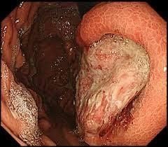
Một ổ loét dạ dày to, đang chảy máu thành dòng, rất khó để can thiệp thành công với các phương pháp cầm máu thông thường
2.3. Stomach cancer
The image of the stomach on X-ray film is also divided into 3 types: lump / lump, solid infection and ulceration.
Nodular form is a common cancer in the stomach, easily recognized on radiographs by a contrast-enhanced area, jagged edges, unchanged on all radiographs. This form usually occurs in the gastric antrum, if the tumor occupies the whole, the image of the stomach is amputated. It is necessary to distinguish the contrast defect caused by gastric cancer with a lump with the spine pressing on the spine when the patient is lying on the stomach (usually in women), food has not been digested...
Hard infection Possible occurs in one region of the curvature or the entire stomach. Stiffness occurring in a segment of the curvature will show the curvature without peristaltic waves; in one area will cause narrowing of the gastric lumen (crystal cancer); In the body area, the image of a double-vesicle stomach will be created, the shape of an X is different from the letter B in a small curvature ulcer.
It is necessary to distinguish gastric cancer with cricoid sclerosis in the antrum (two curvatures no longer have peristaltic waves, loss of mucosal folds) from antral stenosis due to sclerosing inflammation (peristaltic waves of the two gastric curvatures are also visible). and mucosal folds).
Ulcerative and ulcerative cancers Only ulcerative and ulcerative cancers can be identified based on progression of gastric disease. X-ray images are ulcers with small halo-shaped edges (lens or root), mucosal folds around the ulcer or at the bottom of the ulcer that are no longer visible or change direction and fundus. jagged or flat. In fact, a patient may have a combination of the above types of gastric cancer.
Cancer in the upper part of the stomach This type is rare, the lesion is located in the center of gastric aneurysm. X-ray of the stomach will show signs such as: a small defect in the gastric aneurysm, a change in the angle caused by the esophagus entering the cardia, narrowing of the lumen and jagged edges of the esophagus (invasive cancer), signs of Hammer of transcardiac contrast is like water sliding through a fissure in a rock.
Hình ảnh ung thư dạ dày
2.4. pyloric stenosis of the stomach
The common causes of gastric pyloric stenosis are:
Cystic ulcer of the bulb of the duodenum: The stomach is often dilated, lying low, the two curvatures are weak or absent. Circulation of contrast medium into the small intestine is very slow. The stomach contains a lot of food and gastric juice, taking contrast will show signs of snowfall. Stomach cancer: The stomach is less dilated, the contrast medium can still circulate into the duodenum, albeit a bit slowly. In the antrum in the antrum, the gastric lumen will be narrow and stiff; Gastric cancer with gastric nodules in the antrum often creates the image of an amputated stomach. Hypertrophy of the pyloric sphincter. Note, the image of the stomach on the X-ray film is inside the lumen. If you want to evaluate the stomach wall and surrounding organs, it is necessary to add methods such as ultrasound or computed tomography, endoscopic diagnosis of gastric pathology. Although gastric X-ray does not pose any health risks, patients should still conduct it in a room with medical equipment that must meet quality standards and a team of highly skilled technicians to ensure safety. and efficient.
Vinmec International General Hospital has applied X-ray technique in examination and diagnosis of stomach diseases. Accordingly, the stomach X-ray technique at Vinmec is performed methodically and according to standard procedures by a team of highly qualified medical professionals, modern machinery system, thus giving accurate results. contribute significantly to the diagnosis and stage of the disease.
Please dial HOTLINE for more information or register for an appointment HERE. Download MyVinmec app to make appointments faster and to manage your bookings easily.