This is an automatically translated article.
Necrotizing pneumonia in children is an infection of the lung parenchyma, often causing symptoms of cough, fever, and difficulty breathing. The treatment of necrotizing pneumonia is often long and difficult, especially the treatment in children is often more severe than in adults.1. Necrotizing pneumonia in children
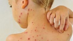
When the weather changes erratically, children are most susceptible to diseases related to lung diseases, especially adding other factors that make children more susceptible to necrotizing pneumonia. The disease is one of the leading causes of death in both adults and children.Necrotizing pneumonia is a severe form of lung disease, caused by infection of the lung parenchyma, causing fever, cough, and difficulty breathing. The disease develops with the formation of granules, small abscesses less than 2cm in the lung parenchyma. The disease has a complicated course and prolonged treatment, especially in young children. The main causes of necrotizing pneumonia are bacteria, viruses, fungi, and parasites. However, depending on age, the causative agent will change.
Nowadays, with the development of antibiotics, necrotizing pneumonia is showing rare signs, but parents should note the following to detect early signs of the disease and take their baby to see them. doctor for prompt treatment.
2. Causes and complications of necrotizing pneumonia in children
2.1 Causes Pulmonary and non-infectious pulmonary infections are the major causes of necrotizing pneumonia. Specifically:Infectious causes: The infectious agents that cause necrotizing pneumonia are: bacteria, fungi, viruses. S. pneumonia, especially pneumococcal strains that are not protected by current pneumococcal vaccines, especially serotypes 3 and 19A are the main causes.
S. aureus, especially the group of methicillin-resistant staphylococcus (MRSA) is one of the causes of necrotizing pneumonia in children. S. aureus produces the toxin Panton-Valentin Leucocidin (PVL) with leukocytosis activity, about 2% of S.aureus strains secrete PVL. In the case of close contact, the percentage of carriers of S.aureus secreting PVL may be higher.
PVL-associated staphylococcal pneumonia has been reported worldwide, with mortality rates ranging from 56-63%, and possibly up to 75%. PVL is a toxin with 2 components LukS-PV and LukF-PV. Both of these components are secreted before consolidating in the heptamers to create small holes in the neutrophil membrane, leading to cell lysis. PVL causes leukocyte breakdown by forming small holes in cell membranes leading to the release of small cytotoxic particles that cause inflammation and lung damage. Accelerated apoptosis, local vasodilation, and tissue necrosis. This phenomenon may even be self-amplifying.
In addition, there are causes such as: inappropriate antibiotic treatment (delayed or incorrect), bacterial virulence or over-response of the patient. But perhaps due to a combination of many factors at the same time.
Non-infectious Nguyen: affected by external factors such as: inhalation of food, chemicals (hydrocarbon, oil, varnish, turpentine), smoke, foreign objects; meconium aspiration syndrome; chemotherapy (Bleomycin, Cyclophosphamide); toxic shock syndrome; rejection reactions; Crohn's disease; systemic lupus erythematosus; psoriasis... 2.2 Complications Necrotizing pneumonia in children, if detected early, will reduce mortality. On the contrary, the disease will have very dangerous complications such as:
Severe infections: septic shock, disseminated intravascular coagulation, acute renal failure, electrolyte disturbances, ARDS. Pleural effusion/emphysema, pneumothorax, air cyst, broncho-pleural fistula, lung abscess. There are reports showing that necrotizing pneumonia with pleural effusion has a very high rate, 80 - 90% with 15 - 55% of broncho-pleural fistulas.

Viêm phổi hoại tử sẽ không nguy hiểm nếu được phát hiện sớm sẽ làm giảm tỷ lệ tử vong
3. Diagnosis of necrotizing pneumonia in children
Through modern diagnostic methods such as X-ray, chest ultrasound, chest CT, the doctor will diagnose whether the patient has necrotizing pneumonia? When pneumonia does not respond to appropriate treatment but requires imaging to confirm the diagnosis, the doctor will prescribe the diagnosis of necrotizing pneumonia.Chest X-ray is one of the commonly used methods of diagnosing necrotizing pneumonia, but it can miss necrotizing pneumonia. The reason is X-ray, the rate of air bubbles seen on chest X-ray is not high (about 22%). This diagnostic method allows the visualization of a pulmonary consolidation lesion with small air bubbles.
Chest ultrasound is a non-invasive method for early detection of necrotizing pneumonia in children, but its role and value in the diagnosis of necrotizing pneumonia are not well defined.
Currently, in the diagnostic method, chest CT is a useful and sensitive method to help confirm the diagnosis of necrotizing pneumonia and assess the spread of the disease. Most studies confirm the diagnosis by chest CT. In addition, chest CT also helps to detect congenital abnormalities or missed airway foreign bodies, if present. This diagnostic method allows to show:
There is an area of pulmonary consolidation but no hypovolemia. Destruction of normal lung parenchymal structures in the presence of areas of reduced contrast enhancement. There are many small gas or fluid-filled cysts (<2cm) (In a lung abscess, the size of the necrotic foci is more than 2cm, the wall thickness is > 2mm).
4. Treatment methods

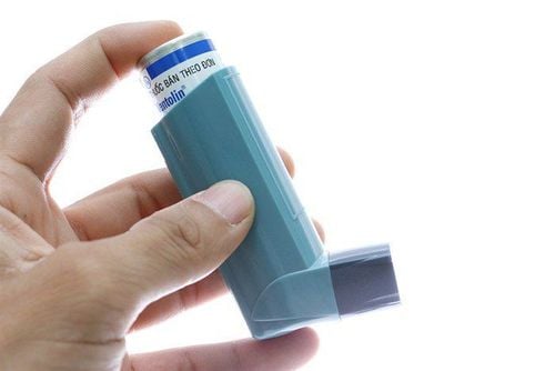
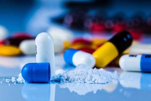
Kháng sinh có tác dụng lớn trong điều trị viêm phổi hoại tử, nhưng cần điều trị kháng sinh theo kinh nghiệm
5. How to prevent necrotizing pneumonia

Cung cấp đủ dinh dưỡng cho trẻ thông qua chế độ ăn uống
Prevention of necrotizing pneumonia is the only way to help children avoid this dangerous disease. Therefore, parents should note some of the following measures to be able to take care of and prevent necrotizing pneumonia for children:
Nutrition, providing enough water for the baby through milk, direct drinking water, porridge , soup... Parents, please encourage children to drink a lot of water (for young children, they should drink small sips of water), give them a reasonable rest. Monitor your baby's urination to see if the water supply is enough. If your baby is urinating less, yellow urine may be due to lack of water. Nasal hygiene: Often children with pneumonia also have upper respiratory infections, can clean the child's nose with physiological saline, or spray mist, remove nasal mucus with a deep wick. Humidify the air in the room. support respiratory mucosa, avoid air-conditioner at too low temperature. When the child has pneumonia, absolutely do not give the child arbitrary medicine. This will make the child's condition worse. Parents, please take your child to the hospital to receive the best support. Washing your child's hands often with soap and water is the best way to prevent the spread of infection. You can also use alcohol-based hand sanitizer instead, but make sure to wash all of your hands. At the same time, parents should go to medical facilities for vaccination to help children avoid necrotizing pneumonia such as: pneumococcal vaccination; vaccination against Hib, pertussis; measles; flu,... Isolate children from cigarette smoke, protect children's respiratory tract, do not allow contact with people who are showing respiratory symptoms. Because there are many dangerous complications, which can lead to death, it is an important step for children to go to the doctor in time to detect necrotizing pneumonia. The Pediatrics Department at Vinmec International General Hospital is the address for receiving and examining diseases that infants and young children are susceptible to: Pneumonia, viral fever, bacterial fever, otitis media, etc. ... Vinmec has a team of experienced pediatric experts in examining, treating and consulting respiratory diseases in children. With modern equipment, sterile space, minimizing the impact as well as the risk of disease spread and professional care services, parents are completely assured when bringing their children to the clinic for examination and treatment. Vinmec.
Please dial HOTLINE for more information or register for an appointment HERE. Download MyVinmec app to make appointments faster and to manage your bookings easily.