This is an automatically translated article.
The article is professionally consulted by Master, Doctor Nguyen Le Thao Tram - Radiologist - Radiology Department - Vinmec Nha Trang International General Hospital.Placenta plays an important role in nurturing the fetus. To ensure the health of the fetus, in addition to ultrasound, placental magnetic resonance imaging is indicated to check the diagnosis and detect some diseases before birth.
1. What is the use of magnetic resonance imaging?
Pregnancy ultrasound is an important method to check the health and development of the fetus. Pregnancy ultrasound includes placental ultrasound. However, for some diseases, ultrasonography does not allow a complete assessment of the status of the placenta. Therefore, magnetic resonance imaging is applied to check the structural and morphological features of the placenta, the results of magnetic resonance imaging are used to supplement the ultrasound results to diagnose and detect some diseases. prenatal illness.

Chụp cộng hưởng từ bổ sung kết quả siêu âm trước khi sinh
2. Indications / Contraindications to magnetic resonance imaging assessment of the placenta in which case?
Placental magnetic resonance imaging is indicated for all suspected placental pathologies detected on ultrasound. Relatively absolute contraindications for pregnant women who have ever had metal surgical clips (over 6 months) or women with agoraphobia. Absolutely contraindicated for pregnant women who use electronic devices such as defibrillators, cochlear implants, ... or have ever had metal vascular surgery, eye socket, intracranial (lower) 6 months), or pregnant women with severe illness must use resuscitation equipment next to them.
3. Steps to perform placental magnetic resonance imaging
For placental magnetic resonance imaging, pregnant women do not need to fast, however, it is important to check for contraindications before the scan.
The imaging procedure includes the following steps:
Step 1: Place the patient supine on the imaging table, select and locate the receiver coil, then move the table into the magnetic field of the scanner, locate the area to capture. Step 2: After taking positioning, the technique consists of 6 pulse sequences. Pulse sequence 1 - HASTE (with breath holding), number of layers performed 24 - 29, thickness of each layer 4 - 5 mm. Pulse sequence 2 - Haste T2 and HASTE FS (with breath hold), image ranges from right margin of uterus to end of left margin, number of layers performed 24 - 29, thickness each layer 5-6 mm. Pulse sequence 3 - Haste T2 transverse scan (with breath holding), perpendicular to the placenta , image ranges from the upper edge of the placenta to the lower edge of the placenta, 4 mm thickness each, possibly Take 2 shots if the shooting time is too long. Pulse series 4 - transverse T1 inphase (with breath holding), image acquisition within the same range as pulse sequence 3, number of layers performed 24 - 29, thickness of each layer 4 mm. Pulse sequence 5 - transverse T1W out of phase (with breath holding), imaging within the same range as pulse sequence 2 HASTE, number of layers performed 24-29, thickness of each layer 5.5mm. Pulse series 6 - cross section HASTE FS is horizontal, following the uterine axis, taking images in the range from the anterior to posterior border of the uterus, the number of layers performed 24 - 29, the thickness of each layer is 6mm. Step 3: At the end of the placental magnetic resonance imaging procedure, the technician prints the film and transfers the resulting data and images to the doctor. The image obtained should show the placenta and its relationship to the uterine muscles, as well as the fetus. Based on images and perfusion parameters, the doctor will analyze and make a diagnosis.

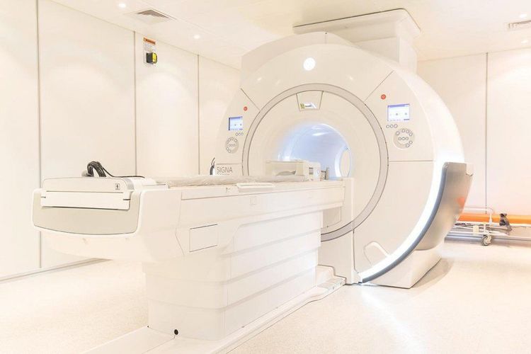
Máy chụp cộng hưởng từ bánh nhau
Placental magnetic resonance imaging is a method that provides image results and data, complementing the ultrasound method in diagnosis and detection of placental pathology.
Currently, Vinmec International General Hospital has put modern machinery and equipment such as magnetic resonance imaging (MRI), computed tomography (CT), X-ray, .. into medical examination and treatment, diagnostic imaging. In particular, the technicians who perform these techniques are all well-trained and qualified medical staff who will give the most accurate image results combined with ultrasound results to offer the optimal treatment regimen for customers.
For detailed advice on this method, please come directly to Vinmec Health System or book online HERE.


