This is an automatically translated article.
This article was written by Specialist Doctor II Khong Tien Dat, Doctor of Radiology - Department of Diagnostic Imaging - Vinmec Ha Long International Hospital.Magnetic resonance angiography of the thoracic aorta is an imaging tool that is increasingly being applied thanks to its outstanding advantages such as clear, detailed images, no radiation exposure. However, because of the relatively long time to perform, MRI of the aorta is not suitable in acute pathology and cannot replace the role of aortic angiography with CT scan.
1. What is the role of magnetic resonance imaging (MRI) of the thoracic aorta?
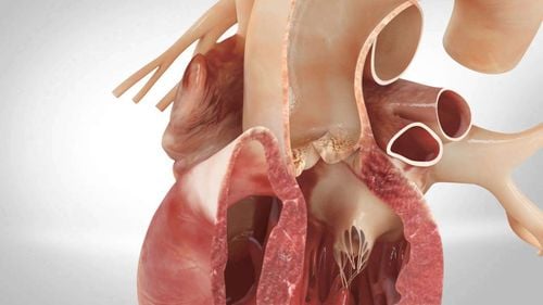
The thoracic aorta is the first part of the aorta and is divided into three segments: the ascending aorta, the aortic arch, and the descending aorta. The thoracic aorta originates from the aortic valve, lies to the left of the midline, corresponding to the 3rd intercostal space. The thoracic aorta is the body's large artery, responsible for supplying blood to the rib cage and extremities. upper, and head and neck areas.Thoracic aortic magnetic resonance imaging or thoracic aorta MRI is a modern, non-invasive, non-X-ray exposure imaging tool with high safety. The role of MRI of the thoracic aorta is well established in the monitoring of aortic pathologies such as aortic dissection, aneurysm mass, atherosclerotic stenosis or spasm. The most commonly used means of assessing the structure and function of the thoracic aorta is Doppler ultrasound. However, Doppler ultrasound of the aorta has many limitations due to its susceptibility to interference by other surrounding structures, as well as the difficulty in assessing the overall morphology. CT scan of the thoracic aorta overcomes the weaknesses of Doppler ultrasound of the arteries, giving a clearer image, but puts the patient at risk of X-ray exposure.
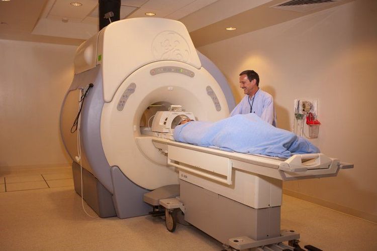
Chụp MRI động mạch chủ ngực là phương tiện chẩn đoán hình ảnh hiện đại
2. Indications for thoracic aortic magnetic resonance imaging in which cases?
Magnetic resonance angiography of the thoracic aorta is indicated in thoracic aortic pathology for the purpose of diagnosis and coordination of treatment planning. However, because of the time-consuming procedure, MRI of the thoracic aorta has not been able to truly replace the role of computed tomography in the acute setting, especially in small centers. Thoracic aortic MRI is usually preferred when the patient is stable,Indications for thoracic aorta MRI include:
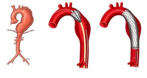
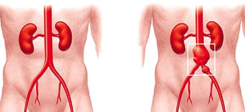
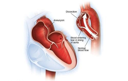
Congenital thoracic coarctation Aortic inflammation Thoracic aortic aneurysmectomy Thoracic aortic dissection Thoracic aortic atherosclerosis Anatomical abnormalities of the aortic arch
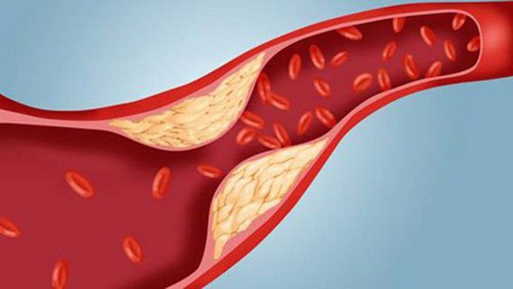
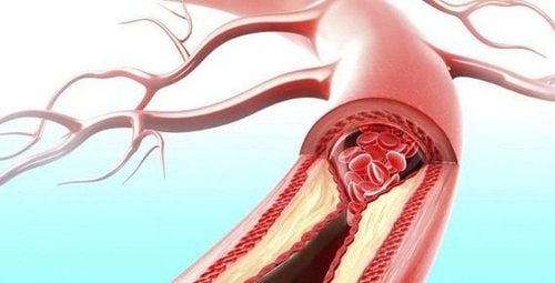
Bệnh nhân xơ vữa động mạch chủ ngực cần được chụp MRI
3. Contraindications for thoracic aorta magnetic resonance imaging in which case?
Magnetic resonance is a safe and highly effective non-invasive means. However, MRI of the thoracic aorta is not always permitted.Contraindications to thoracic aortic magnetic resonance imaging include:
People with claustrophobia: The magnetic resonance imaging process requires the patient to lie still in a position under the imaging chamber for a period of time. People with claustrophobia may not cooperate to produce quality images. In some cases, the doctor may have to prescribe sedation. Patients bring foreign objects or replacement materials containing iron, with magnetism such as pacemakers, artificial cochlea, artificial heart valves, bone fusion facilities, vascular disease treatment clips, ... Magnetic waves can damage these devices and affect the health of the patient. Patients with metal foreign bodies in the eye, or shrapnel in the body are also not in the indications for MRI of the thoracic aorta.
4. What should the patient prepare before thoracic aorta?
Some notes that patients need to remember before conducting MRI of the thoracic aorta:Patients do not need to fast if the MRI is taken without drugs. With contrast-enhanced MRI, the patient needs to remember not to eat or drink anything 4 hours before. Do not bring jewelry, earrings or anything made of metal such as watches, bank cards. Medicines taken daily to treat chronic diseases such as high blood pressure and diabetes can still be used normally as prescribed by the doctor. Declare detailed medical history, including surgical history of implantation of artificial materials to detect contraindications if any.

Trước khi chụp MRI, bệnh nhân không cần nhịn ăn
5. Steps to perform a thoracic aortic angiogram
MRI of the thoracic aorta should be performed according to the correct procedure to ensure patient safety as well as ensure the best image quality.The steps to perform a thoracic aortic angiogram include:
Prepare the magnetic resonance machine and basic tools Advise and clearly explain to the patient the steps in the magnetic resonance imaging procedure. Exploit detailed history and medical history to rule out contraindications. Ask the patient to remove jewelry and personal items, change clothes as prescribed before entering the MRI room. Family members accompanying the patient are also not allowed to bring metal objects into the examination room. Instruct the patient to lie on his back in a chair, tie the breathing belt around waist level, not too tight or too loose. Instruct the patient to follow the technician's instructions. The MRI scan generates a lot of noise, so it is necessary to equip the patient with noise-reducing means such as earplugs or noise-canceling headphones. The total time for a single shot can be up to 1 hour. Vinmec International General Hospital with a system of modern facilities, medical equipment and a team of experts and doctors with many years of experience in neurological examination and treatment, patients can completely rest in peace. Center for examination and treatment at the Hospital.
With more than 14 years of working in the field of BSCK II imaging, Khong Tien Dat is currently a radiologist at Vinmec Ha Long International Hospital.
To register for examination and treatment at Vinmec International General Hospital, you can contact Vinmec Health System nationwide, or register online HERE.


