This is an automatically translated article.
The article was professionally consulted by resident Doctor Nguyen Quynh Giang - Department of Diagnostic Imaging and Nuclear Medicine - Vinmec Times City International Hospital.The shoulder joint is an important joint during movement. The shoulder joint has a greater range of motion than other joints. Magnetic resonance imaging (MRI) is considered the best diagnostic method for diseases related to bones and joints, including shoulder joints.
1. Magnetic resonance imaging for what?
Magnetic Resonance Imaging (MRI) is one of the revolutionary inventions in the field of medical engineering. Instead of using X-rays, with the principle of magnetic fields, MRI gives clearer images, allowing the ability to reproduce images in three-dimensional space. Thereby, helping to diagnose more accurately, playing a decisive role in treatment in a variety of diseases. Magnetic resonance imaging has a wide range of applications and is safe and does not cause radiation to the patient.Magnetic resonance imaging with high contrast, good anatomical details allows accurate detection of morphological and structural lesions in body parts.
The ability to reproduce 3D images, without side effects, is increasingly being specified for many different specialized applications. It has important applications in the diagnosis and treatment of musculoskeletal diseases.
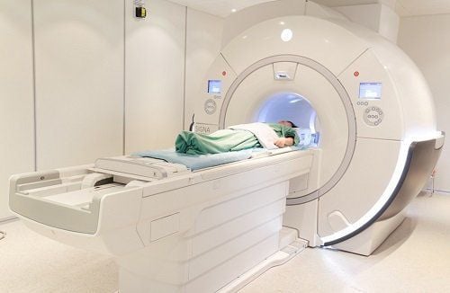
Chụp cộng hưởng từ MRI (Magnetic Resonance Imaging) là một trong những phát minh có tính cách mạng trong lĩnh vực kỹ thuật y học
2. Shoulder joint magnetic resonance imaging
The shoulder joint is an important joint in movement. The shoulder joint has a greater range of motion than other joints. However, the shoulder joint is not as stable as other joints because the surface of the socket is small and shallow relative to the humerus head. The shoulder joint involves many bony and soft structures around the joint. Anatomical or pathological changes to these structures affect joint mobility.MRI of the shoulder joint is the best imaging method of musculoskeletal system to comprehensively evaluate the structures of the joint including: shoulder arthritis, bone damage, cartilage, bursitis, ligaments, tendons and muscles as well as soft tissue organization around the joint that other imaging methods cannot do.
2.1. Indications for magnetic resonance imaging of the shoulder joint ● Abnormalities, injuries to the rotator cuff, tendons, ligaments and cartilage, dislocations and fractures.
● Capture after injury due to vigorous activity, playing sports: Ligament injury, ligament tear, tendon tear, ...
● Detect fracture when not visible on X-ray image or on tomography computer and other diagnostic imaging.
● Inflammatory and degenerative conditions such as: Osteomyelitis, osteoarthritis
● When there is a feeling that the humerus head is not connected in the joint socket
Disease of the bursa, shoulder muscles, tumors and shoulder arthritis.
● Monitor complications, progress after intervention, joint surgery.
Disorders of bone marrow, nerves, blood vessels.
2.2 Contraindications to shoulder magnetic resonance imaging ● Patients with cardiovascular support equipment such as pacemakers, defibrillators, prosthetic heart valves...
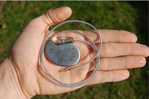
Chống chỉ định chụp cộng hưởng từ khớp vai với bệnh nahan có thiết bị hỗ trợ tim mạch như máy tạo nhịp tim
● People with claustrophobia.
● People who are obese, whose body weight is too large to fit the cage of the magnetic resonance machine or the receiving coil.
3. Uses of shoulder joint MRI
Compared with other joints, the shoulder joint has greater range of motion but is not as stable as the socket surface is smaller and shallower than the humerus head. Many of the bony and soft structures around the joint are involved in the shoulder joint. These anatomical or pathological changes associated with this structure affect joint mobility.MRI of the shoulder joint can detect bone lesions in trauma such as non-displaced fractures, contusion of bone marrow edema, trauma cases where X-ray or electronic tomography have not been detected. Not only that, bone and joint magnetic resonance imaging also helps to evaluate soft shoulder joint injuries, muscle tendons, ligaments, etc. associated with bone injuries.
Vinmec International General Hospital put into use the magnetic resonance imaging machine 3.0 Tesla Silent technology. Magnetic resonance imaging machine 3.0 Tesla with Silent technology of GE Healthcare (USA).
● Silent technology is especially beneficial for patients who are children, the elderly, weak health patients and patients undergoing surgery
● Limit noise, create comfort and reduce stress customers during the shooting process, helping to capture better quality images and shorten the shooting time.
● Magnetic resonance imaging technology is the technology applied in today's most popular and safest imaging method because of its accuracy, non-invasiveness and non-X-ray use. many years of experience in the field of diagnostic imaging, especially in the field of multi-segment computed tomography, magnetic resonance. Currently, the doctor is working at the Department of Diagnostic Imaging and Nuclear Medicine - Vinmec Times City International Hospital.
Please dial HOTLINE for more information or register for an appointment HERE. Download MyVinmec app to make appointments faster and to manage your bookings easily.