This is an automatically translated article.
The article is professionally consulted by Master, Doctor Ton Nu Tra My - Department of Diagnostic Imaging - Vinmec Central Park International General Hospital.Magnetic resonance imaging of Parkinson's disease plays an important role in the differential diagnosis of Parkinson's disease and some pathologies that cause Parkinson's syndrome. From these diagnostic results, doctors can distinguish, determine the stage of the disease and give appropriate treatment.
1. Features of Parkinson's disease
Parkinson's disease is a degenerative form of the central nervous system that affects a person's ability to move, balance, and control muscles. Parkinson's disease is common in the elderly, the older you are, the greater the risk of developing the disease. Both men and women can develop Parkinson's disease, but the incidence is higher in men than women. Scientists have not yet found a specific cause of Parkinson's disease, but many studies have shown that the level of Dopamine in the body of patients is significantly reduced. Dopamine is a neurotransmitter that plays an important role in transmitting signals between nerve fibers in the brain, helping with movement and coordination of body movements. When brain cells are degenerated, the amount of Dopamine in the patient's body will be reduced, making it difficult for the patient to move.In the early stages of Parkinson's disease, people with Parkinson's disease may have symptoms such as:
Fatigue, muscle pain, constipation, difficulty in performing simple tasks such as wearing shoes, sandals,... Reduced one-handed activity when exercising, writing disorders, sweating and scaly skin on the knees A small number of patients have symptoms of tremor at rest but intermittently and discreetly.

Tremor: is a symptom clearly seen in the lips, tongue, and extremities. Tremor usually occurs on one side of the body for the first few years, after a few years can progress to bilateral tremor. The side with tremors first also tends to get worse throughout the course of the disease. Stiffness: The patient's limbs are stiff in all muscle groups, causing difficulty in movement. This is one of the most important symptoms. Decreased movement: the patient loses facial expression, blinks less. Loss of natural movements of the limbs, especially when moving. Other common symptoms are edema, cyanosis of the extremities, stomach and bladder dysfunction, orthostatic hypotension, pain sensation, depression, severe dementia, etc.
Need to distinguish Parkinson's disease and Parkinson's syndrome. Parkinson's syndrome includes symptoms similar to Parkinson's disease, but other conditions cause these symptoms.
2. Parkinson's disease magnetic resonance imaging
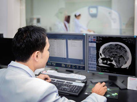
Cranial magnetic resonance imaging is a technique that creates anatomical images of the body using magnetic fields and radio waves. This is a safe, non-invasive method that gives high-definition images, helping to detect brain lesions and abnormalities at an early stage.To diagnose Parkinson's disease, doctors will rely on clinical symptoms and order a number of specialized tests. Magnetic resonance imaging (MRI) of the brain has an important role in the differential diagnosis of Parkinson's disease and some diseases causing Parkinson's syndrome.
Parkinson's disease magnetic resonance imaging has the following features: black matter atrophy in the brain stem. On T2 images, the black crescent can be seen to be less signal-intensive than usual. Cranial magnetic resonance imaging also helps detect the causes of secondary Parkinson's syndrome.

3. What should patients pay attention to when taking magnetic resonance imaging in Parkinson's disease?
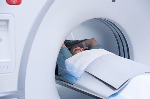
When a doctor appoints cranial magnetic resonance imaging to diagnose Parkinson's disease, the patient should note the following points to ensure safety when taking the scan:Patients are not allowed to bring into the MRI room any items that have dangerous effects on them. metal such as jewelry, watches, cell phones, keys, etc. The high magnetic field of the scanner can damage metal implants in the body. Therefore, the patient needs to inform the radiologist if he or she is using a hearing aid, a pacemaker, an artificial heart valve, etc. Try to stay still while taking the picture to get good quality images. Patients do not need to fast before cranial magnetic resonance imaging. Only in case anesthesia is needed, the doctor will tell the patient to fast for 4-6 hours before the scan. The scan time usually ranges from 15-45 minutes, depending on the complexity of the survey and the cooperation of the patient.

Please dial HOTLINE for more information or register for an appointment HERE. Download MyVinmec app to make appointments faster and to manage your bookings easily.


