This is an automatically translated article.
The article was consulted with Dr. Nguyen Luong Tan - Head of Cardiology Department - Cardiovascular Center - Vinmec Central Park International General Hospital.Atrial septal ablation is performed in the treatment of congenital heart disease by inserting an instrument through a blood vessel in the patient's thigh, bringing the instrument to the heart to block holes in the heart or widen narrow areas in the heart. heart valves or blood vessels.
1. Indications for rupture of the atrial septum
The method of breaking the atrial septum, treating congenital heart disease is indicated in the following cases:Inversion of the artery with an intact interventricular septum, or limited small ventricular septal defect. Abnormal pulmonary veins to the heart completely with atrioventricular septal defect, limited oval foramen, severe pulmonary hypertension. Fulminant pulmonary valve stenosis. Pulmonary valve atrophy with intact interventricular septum. Tricuspid atrophy with limited atrial septal defect. Mitral valve atrophy without being able to perform Norwood surgery. Two-way outflow right ventricle with intact interventricular septum and limited atrial septal defect.
2. Contraindications for rupture of the atrial septum
Contraindications are as follows:Severe coagulation disorders. Having other serious internal or surgical diseases that cannot be catheterized.

Chống chỉ định đối với trường hợp rối loạn quá trình đông máu
3. Methods of tearing the atrial septum
There are 4 types of atrial septal defect encountered clinically. Depending on the type of vent, there will be different treatment directions.First hole atrial septal defect accounts for about 15-20% of all types of atrial septal defect. The first hole is usually located in the lower region of the atrial septum. Second-hole atrial septal defect accounts for about 75% of atrial septal defects. This opening is located in the middle of the atrial septum. This type of atrial septal tear can be completely cured with parachute occlusion through cardiac catheterization. Sinus-venous atrial septal rupture is rarer, accounting for 5-10% of cases. The location of this septum is usually located in the upper part of the atrial septum. The rarest form is a ruptured atrial septum of the coronary sinus with the septum located between the atrial septum and the coronary sinus.
4. Steps to break the atrial septum
4.1. Breaking the atrial septum under the guidance of an angiogram Posture: The patient lies in the supine position, arms raised above the head. Medical staff anesthetize according to the anesthetic procedure. The doctor performed an puncture of the femoral vein. Angiography machine to straighten the face. Break the balloon atrial septum or catheter with the lead from the femoral vein to the inferior vena cava, to the right atrium, through the foramen ovale to the left atrium, if it is a catheter, withdraw the catheter, keep the lead in place, then Insert the atrial septal balloon into the left atrium through the lead, then remove the lead. Inflate the balloon with a syringe that dilutes the contrast medium at a rate of 25%. After determining the ball head in the left atrium, the balloon is inflated and recoiled from the left atrium to the right atrium, the jerking action must be strong enough and sized to avoid pulling the balloon too hard and not The size can cause tearing of the inferior vena cava, conversely, pulling the ball with insufficient force will not widen the foramen ovale. This move is done a few times to make sure the oval hole is enlarged.
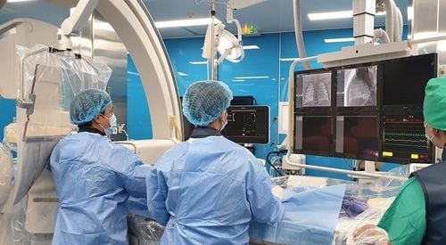
Phá vách liên nhĩ dưới sự hỗ trợ của máy chụp mạch cần được thực hiện tại bệnh viện uy tín
4.2. Break the atrial septum at the emergency bed under ultrasound guidance. The patient lies in the supine position, with the buttocks elevated. The patient is anesthetized with sedation and mechanical ventilation. The doctor punctured the femoral vein. Break the ball of the atrial septum from the femoral vein into the inferior vena cava, to the right atrium, through the foramen ovale to the left atrium under ultrasound guidance. Check where the balloon position is by injecting physiological saline along the path of the balloon (the way into the blood vessels, not the way to inflate the balloon) if you see air bubbles in any position, it is the position of the balloon tip. Inflate the balloon with a syringe of physiological saline solution. After determining the tip of the balloon in the left atrium, inflate the balloon and pull the balloon back from the left atrium to the right atrium, the jerking motion should be strong enough and sized to avoid pulling too hard and not have a suitable size. can cause a tear in the inferior vena cava, conversely, when pulling the ball with insufficient force, it will not widen the foramen ovale. This maneuver is done several times to make sure the foramen ovale is enlarged on echocardiography. At the end of the procedure to break the atrial septum, it is necessary to withdraw the balloon, then withdraw the vascular catheter from the femoral vein, squeeze the femoral vein by hand, monitor until the bleeding stops, then apply pressure with an elastic bandage.

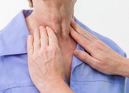
Khám và sàng lọc bệnh tim bẩm sinh là việc làm cần thiết
5. Complications of atrial septal rupture
5.1 Complications during the procedure Pericardial bleeding: Blood transfusion, pericardial aspiration, surgery when necessary. Venous bleeding due to tear: Compression bandages, blood transfusion, surgery when necessary. Cardiac arrhythmias: Treatment of arrhythmias according to each type of arrhythmia, arrhythmia drugs, electric shock... Late complications: Hematoma at femoral vein puncture: Compression bandages, hemostasis stitches... Infarction , embolism: Right heart failure, arrhythmia or stroke. Pulmonary hypertension results from increased blood flow to the lungs during an atrial septal defect and constant pressure on the pulmonary capillaries (pulmonary hypertension). This is the most dangerous late complication of atrial septal rupture. If not treated promptly and correctly, pulmonary hypertension can lead to Eisenmenger syndrome (causing permanent pulmonary hypertension). Atrial septal rupture is an urgent procedure to improve the oxygenation and hemodynamics of the pediatric patient. Immediate examination is needed to confirm the diagnosis of a life-saving procedure strategy, and to prepare for a semi-urgent or elective surgical treatment step.Vinmec has a team of pediatric echocardiography experienced in diagnosis and treatment. There is a Hybride room for both interventional and cardiac surgery without moving the patient.
Please dial HOTLINE for more information or register for an appointment HERE. Download MyVinmec app to make appointments faster and to manage your bookings easily.