This is an automatically translated article.
Dilatation of the posterior fossa in the fetus is a dangerous birth defect, which should be detected and treated early to avoid the risk of hydrocephalus, which greatly affects the health of the fetus and mother.
1. What is the posterior fossa dilatation in the fetus?
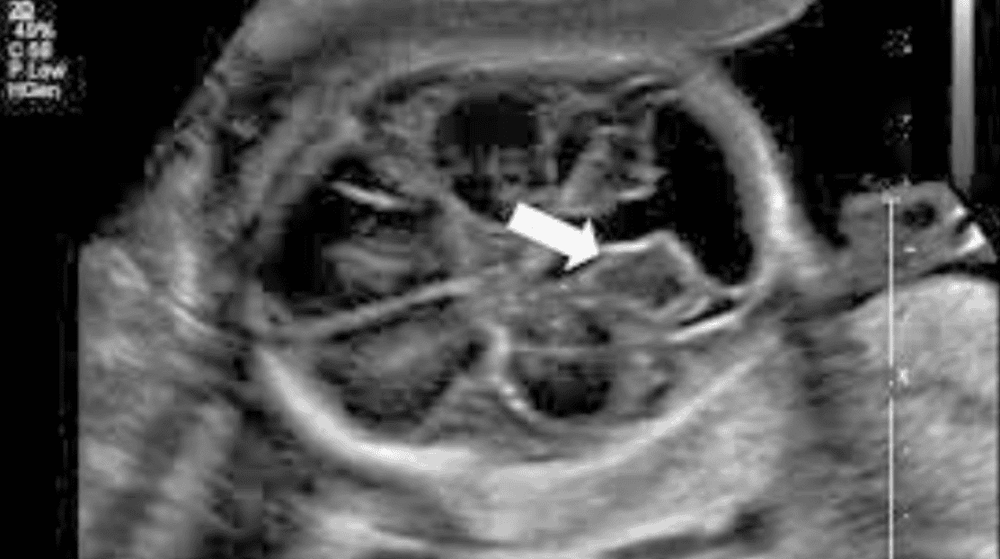
Dilatation of the posterior fossa cistern in the fetus is a condition in which ultrasound results show that the diameter of the posterior fossa cistern is larger than the average posterior fossa cistern diameter according to gestational age.
However, most posterior fossa dilatation is not associated with chromosomal disorders and the prognosis is good. In the case of posterior fossa dilatation but in the setting of Dandy Walker syndrome (including posterior fossa dilatation, cerebellar nystagmus, lateral ventricle dilation), 28% is related to chromosomal disorders and ability 20-30% may be the cause of mental retardation later in life.

Giãn bể lớn hố sau ở thai nhi được phát hiện thông qua siêu âm
2. Types of dilatation of the posterior fossa in the fetus
Large dilatation of the posterior fossa in the fetus is common in the following phenotypes:Blake's pouch cyst. Large tank with simple dilatation of posterior pit (Megacisterna magna). Dandy–Walker syndrome (Dandy–Walker malformation). Vermian hypoplasia of the cerebellum. Cerebellar hypoplasia.
Ultrasound can differentiate these cases with 80% accuracy. With additional MRI, additional information can be obtained with >90% accuracy. It is essential to accurately diagnose the phenotype because each case has a completely different prognosis.
Blake pouch and simple posterior fossa dilation: 30-40% may spontaneously regress during pregnancy. Even in those cases that persist after birth, the prognosis is good with >90% living well without other abnormalities or mental retardation later in life. Particularly, Dandy-Walker syndrome, nystagmus or partial cerebellar hypoplasia, the possibility of associated chromosomal abnormalities and mental retardation and balance and movement disorders later in life is up to 50 years. %.

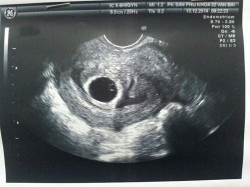
Giãn bể lớn hố sau trên kết quả siêu âm thai nhi
3. Is it dangerous to dilate the posterior fossa in the fetus?
Normal in ultrasound, posterior fossa diameter < 10mm. If > 14mm, the possibility of hydrocephalus is very high.
The posterior fossa is a small space in the skull found near the brainstem and cerebellum. During pregnancy the brain begins to develop from a tubular structure. As the neural tube develops, the inner tube partially forms the spinal cord and partially develops into chambers called the ventricles located in the brain, which communicate with each other.
Inside the ventricles, the arachnoid plexus begins to produce cerebrospinal fluid by the sixth week of pregnancy. CSF will be produced 300-500cc per day in the neonatal period.
The cerebrospinal fluid will pass through many holes in the brain and exit into the subarachnoid space, where it is absorbed into the brain. When there is an obstruction, the cerebrospinal fluid will gradually stagnate, creating dilated ventricles and hydrocephalus with less and less brain tissue due to the dilated ventricles.
On ultrasound, in the second trimester, it is called posterior fossa dilatation if the diameter is over 10mm and hydrocephalus if over 15mm. The brain tissue may not be significantly damaged when ventricular dilatation occurs. But if it progresses, there may be irreversible brain damage.
Hydrocephalus is one of the most common neurological abnormalities, accounting for 0.3-2.5 per 1,000 live births. The treatment of large cisterns after postpartum dilatation is the placement of a catheter connected to the abdominal cavity quite successfully. As for the placement of the ventricular junction - amniotic cavity was abandoned because it was too expensive.
For all information about maternity and childbirth services at Vinmec Hospital, please contact the hospitals and clinics of Vinmec Health system nationwide
Please dial HOTLINE for more information or register for an appointment HERE. Download MyVinmec app to make appointments faster and to manage your bookings easily.


