This is an automatically translated article.
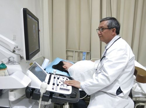
The article was professionally consulted by Doctor Nguyen Thi Mai and Doctor Nguyen Thi Hong On - Department of Obstetrics and Gynecology, Vinmec Phu Quoc International General Hospital.Fetal ultrasound is a technique that has been widely used in the medical field since 1950. This is considered a safe method, does not affect the fetus and brings high diagnostic efficiency, accurate monitoring. the baby's development in the womb.
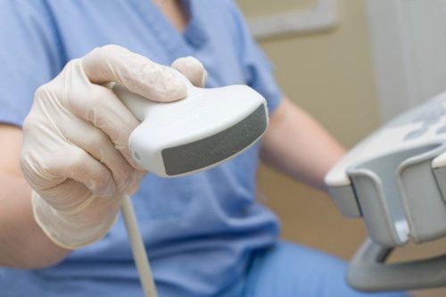
Ultrasound scanners are used by doctors to read important information and provide images of the fetus on a screen, which can also be seen by parents. However, it is difficult for parents to understand the information and ultrasound results displayed on the ultrasound sheet that the doctor did not mention.
1. Meaning of symbols in fetal ultrasound results
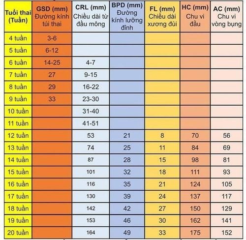
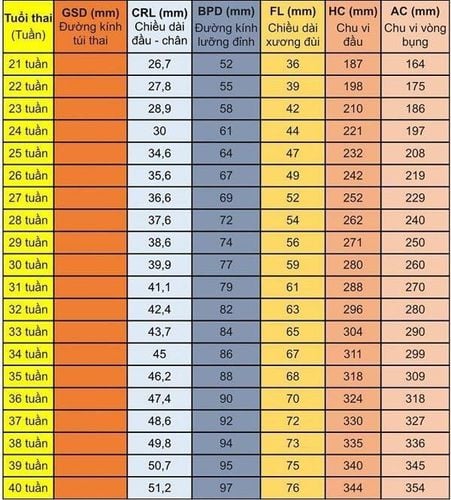
GS: gestational sac TTD: transverse abdominal diameter APTD: anterior and posterior abdominal diameter BPD: biparietal diameter (maximum diameter measured across temporal bone horizontally) OFD: occipital diameter (diameter measured in the largest section - from forehead to nape of fetal skull) CER: cerebellar diameter THD: thoracic diameter AC: abdominal circumference HC: head circumference CRL: rump-head length FL: bone length femur HUM: length of humerus Ulna: length of tibia: length of tibia Radius: length of radial fibula: length of fibula AF: amniotic fluid AFI: amniotic fluid index BD: orbital distance : uterine height. EFW: fetal weight GA: gestational age EDD: estimated date of birth Rump : bottom of baby's bottom. Head position: the baby is in the normal position (head is below). TT(+): fetal heart is heard. TT(-): fetal heart is not heard. Para 0000: a woman who has never given birth (sons). VDRL: test for syphilis. HIV (-): negative AIDS test. CCPT: occipital bone rotates to the right, forwards. CCTT: occipital bone rotates to the left, forwards. CCPS: occipital bone rotates to the right posteriorly. CCTS: occipital bone rotates left posteriorly.2. Fetal index measurement table for reference
3. Important times to have a pregnancy ultrasound
Ultrasound in the first trimester: In the first 3 months of pregnancy, from the 11th to the 13th week, 6 days, the fetal malformation ultrasound at this time is very important:1: Provide basic information of the fetus: Confirm the fetus is still alive or not? See if the fetus is in the right position? How many pregnancy? Calculate the exact gestational age based on the length of the head of the buttocks. Second: Fetal ultrasound during this time is a golden time to detect some fetal abnormalities if any, the time to measure the nape of the neck to predict some chromosomal abnormalities (these abnormalities can be may be: Down disease, heart malformation, ...). In addition, fetal malformation ultrasound during this time also helps detect a number of other fetal malformations such as:
Nervous defects such as: Anencephaly, no anterior division of the brain, spina bifida (manifestations) in the form of hidden spina bifida, meningeal hernia, meningeal-meningeal hernia)... Abnormalities in maxillofacial, lip, eye: Cleft palate, cleft lip-cleft palate... heart and thoracic defects such as: tetralogy of fallot, inversion of the artery, hypoplasia of the left ventricle, thoracic hernias... Abdominal anomalies such as: umbilical hernia... Bone malformations, limbs such as: bone dysplasia, bone hypoplasia, incomplete bone formation, aplasia of cartilage, defects in the number of limbs... In fact, in this stage, the baby has developed relatively fully in terms of morphology. and has reflexes such as flexing and stretching the body, stretching the limbs... This is also one of the 03 important ultrasound landmarks recommended by experts to perform. During the 12-week ultrasound, doctors will especially check and screen for early abnormalities of the brain, face, heart, digestive, urinary, extremities and the whole body.
Because the baby is still quite small, a high-end 4D ultrasound system like the GE Voluson E10 will play a very important role in helping doctors at Vinmec detect more than 95% of anomalies during this period. Packed with the latest technologies, the GE Voluson E10 enables enhanced image quality and penetration for outstanding high-resolution images and easy operation.
Fetal ultrasound at 18 to 23 weeks: At this time, the fetus has basically fully developed its organs and organs, and the amount of amniotic fluid is also increased, allowing a good observation of the fetal morphology. . This is the standard ultrasound time to evaluate the entire fetus.
This is an important milestone to detect most of the morphological abnormalities, confirm the abnormalities that were previously suspected, the final time for the decision to terminate the pregnancy if any (before the 28th week). Most morphological abnormalities can be diagnosed at this stage, the sonographer will look at the fetal parts in turn to evaluate the whole: Neurological abnormalities such as: Abnormalities neural tube, no brain, hydrocephalus, dilated ventricles, baby brain, varicose veins of galen... Abnormalities of maxillofacial: Abnormalities are more clearly observed at ultrasound in the first month, especially Abnormalities in the eye can be observed. Cardiovascular abnormalities: At this stage, fetal ultrasound can clearly observe the heart and its structures, allowing the diagnosis of most abnormalities, including the most complex such as: atrioventricular, tetralogy of fallot, hypoplasia of the heart valves, Ebteins' disease, right ventricular 2 outflow tract, cardiac arrhythmias... Thoracic abnormalities: diaphragmatic hernia, pulmonary cyst, pleural effusion, oliguria Abnormalities in the abdomen, intestines and abdominal wall such as: esophageal stenosis, gastric stenosis, hepatomegaly, splenomegaly, intestinal obstruction, umbilical hernia.... Abnormalities in the kidneys and urinary tract such as : Absence of kidney, polycystic kidney, urinary tract obstruction, abnormalities in bladder, urethra... Abnormalities in skeletal muscle and extremities: In addition to abnormalities detected in the first trimester ultrasound, stage This section observes in more detail the fingers and feet, and it is easy to detect defects such as many fingers, crooked hands...

Ultrasound in the last 3 months: Week 30 - 32:
This is the stage when the fetus has fully completed its structure, and develops rapidly. Fetal malformation ultrasound at this stage is mainly to evaluate fetal growth, fetal position, amniotic fluid, umbilical cord (and their abnormalities if any), the development of the uterus.. Fetal abnormalities that may be further detected or evaluated at this stage than in the mid-month period) include: Fetal malnutrition, reproductive system abnormalities (sperm position and movement). testicles, genital tumors, ovarian cysts...) , some abnormalities in the heart valves were observed more fully (heart tumor, stenosis of the heart valves, mitral aortic valve, aortic abnormalities...), some brain abnormalities. Choosing a location for maternity care and fetal ultrasound to diagnose fetal malformations early and accurately is very important. It is very important to have a pregnancy ultrasound at the right time and periodically to detect fetal malformations early, so that appropriate monitoring and treatment measures can be taken (even a decision to terminate the pregnancy). The effectiveness of the ultrasound method for diagnosing fetal malformations depends a lot on the qualifications of doctors and modern equipment.
Vinmec International General Hospital is a high-quality medical facility that is highly appreciated by customers for its quality of examination and treatment as well as professional services. Vinmec has the most modern equipment system, with the most advanced ultrasound machines in the world and a team of experienced obstetricians in prenatal diagnosis and intervention to help monitor and detect early birth defects.
In addition, to protect the health of mother and baby comprehensively, Vinmec offers a variety of package Maternity services. With this package, pregnant women will have regular antenatal check-ups with specialist doctors, especially thyroid screening tests for mothers in the first 3 months, Rubella tests. Perform prenatal and postnatal screening tests for the baby. During pregnancy, if a pregnant woman encounters any abnormal health problems, she will be consulted and treated by a specialist doctor. After each visit, the doctors will analyze the ultrasound and test results and advise on health care, nutrition, and rest suitable for each pregnant woman, helping the mother and fetus to be healthy. well developed, uniform indicators.

Please dial HOTLINE for more information or register for an appointment HERE. Download MyVinmec app to make appointments faster and to manage your bookings easily.


