This is an automatically translated article.
The article is professionally consulted by Dr., Dr. Tran Nhu Tu - Dean and Master, Doctor Nguyen Van Huong - Department of Diagnostic Imaging - Vinmec Danang International HospitalMagnetic resonance imaging of the skull base and stony bones not only helps doctors to recognize many diseases early, but also evaluates localized or diffused lesions, focusing on the base of the skull.
1. Magnetic resonance imaging of the base of the skull and bones
Magnetic resonance imaging is a method of taking pictures of organs in a living body and assessing the amount of water inside the structure of the organs, using magnetic fields and radio waves. This method does not use X-rays, so it is completely harmless to health.
Accordingly, the magnetic resonance imaging of the skull base and the stony bones is to evaluate the focal or diffuse lesions, which are concentrated in the skull base or the stony bones.
However, magnetic resonance imaging may present some difficulties in cases where the patient is young, fussy, or who has contraindications to MRI.

Cấu tạo nền xọ
2. Indications and contraindications for magnetic resonance imaging of the skull base and stony bones
2.1 Indications Examination of diseases of the skull base and stony bones Infection, inflammation Tumor lesions 2.2 Contraindications Absolute contraindications:
Patients wearing electronic devices such as pacemakers, anti-vibration machines, cochlear implants, automatic subcutaneous injection devices, Neurostimulator...are not allowed to enter the magnetic resonance imaging room. Intracranial metal objects, eye sockets, blood vessels < 6 months Severely ill patients needing resuscitation equipment next to the body Relative contraindications:
Metal surgical forceps > 6 months Patients with fear of the dark, afraid of being alone

Chống chỉ định đối với người có cấy ghép ốc tai điện tử
3. Steps to perform magnetic resonance imaging of skull and stony bones
3.1 Preparation To perform magnetic resonance imaging of the skull and bones requires facilities such as: magnetic resonance imaging machine, specialized electric pump, and image storage system, film and film printer.
Some of the drugs used include: contrast agents, sedatives, skin and mucosal antiseptics.
Before performing magnetic resonance imaging of the skull base and stony bones, the patient should be checked to ensure the safety of magnetic resonance, carefully explain the procedure to coordinate with the doctor, check the contraindications Patients do not need to fast. The medical staff instructs the patient to change into the clothes of the MRI room and remove contraindicated items.
3.2 Steps to conduct magnetic resonance imaging of the skull and scapula Place the patient
The patient is lying supine on the table Select and position the receiver Move the table into the machine's magnetic field and locate the area take a shot
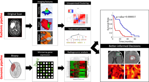
Chụp cộng hưởng từ nền sọ và xương đá giúp đánh giá mức độ tổn thương
Capture basic pulse sequences before drug injection
Transverse T2 localization, 3 mm. Can be replaced or supplemented with high-resolution 3D pulse sequences such as CISS 3D T2 frontal plane, 3mm, can be used with or without transverse T1, 3mm. Diffusion echo-planar- imaging or Diffusion HASTE, section thickness 3 - 4mm, in cross-section. Photograph after drug injection
Need to inject contrast agent if necessary at a dose of 0.1mmol/kg of body weight at a rate of 2ml/sec. T1 cross section 3mm, with fat removal T1 frontal plane 3mm, with fat removal. Can be replaced by 3D T1 pulse sequence with fat removal. The technician will process images, print film, transfer images and data to the doctor's work station. Doctor analyze diagnostic images. Therefore, they are indicated in the examination of cranial and stony-bone pathologies, inflammation, infection, or tumoral lesions. When the patient has an injury or abnormal symptoms, it is necessary to immediately go to a medical facility for a timely examination and diagnosis.

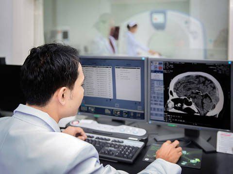
Kết quả chụp MRI nền xọ
Vinmec International General Hospital is the first unit in Southeast Asia to put into use the new 3.0 Tesla Silent MRI machine manufactured by GE Healthcare USA.
The machine currently applies the safest and most accurate magnetic resonance imaging technology available today, without using X-rays, non-invasive. Silent technology reduces the discomfort caused by noise, which is very good for patients who are young children, the elderly, patients with weak health or have just had surgery.
To evaluate the stony bones requires a CT scan to capture very thin slices and many slices for multi-plane reconstruction, with the 640-slice CT at Vinmec this is done quite easily for the patient. images with very high resolution. Moreover, the hospital's MRI is capable of capturing 3D image precochlear system for the diagnosis of abnormalities in parts of the middle and inner ear very accurately.
Please dial HOTLINE for more information or register for an appointment HERE. Download MyVinmec app to make appointments faster and to manage your bookings easily.