This is an automatically translated article.
The stenvers position x-ray technique aims to expose the anterior surface of the scapula, the lower 2/3 of the mastoid bone, and the posterior part of the occipital bone to evaluate the lesions of the mastoid and scapula.Stenvers position x-ray is often used to diagnose and study traumatic brain injuries that cause fractures along the transverse contour, osteomyelitis, and tumors in the cerebellar pons. Cases of suspected stone fracture, damage to the semicircular canals, at the top of the stony bones, and the inner ear canal should also be indicated for stenvers position x-ray.
Thanks to X-ray stenvers position, the doctor can see on film the parts of the cochlea, inner ear, inner ear canal, vestibule, superior and outer semicircular canal, and the stony process. From there, it is diagnosed that there is a bone fracture line, the patient has a broken stone bone, the inner ear canal is elastic, which can be a sign of nerve tumor VIII, diagnose sinus cavity diseases, support in the treatment of extreme for the patient.
1. Prepare for stenvers . x-ray
Instruments such as x-ray machine, right and left marks, doctor's order card are fully prepared and stabilized before taking pictures. The doctor or technician will perform the stenvers position x-ray for the patient.
Patient must remove earrings, necklace, hairpin... if any before being invited into the imaging room with detailed explanations and necessary instructions during the x-ray process.

Kỹ thuật chụp x quang tư thế stenvers
2. Conduct stenvers . x-ray
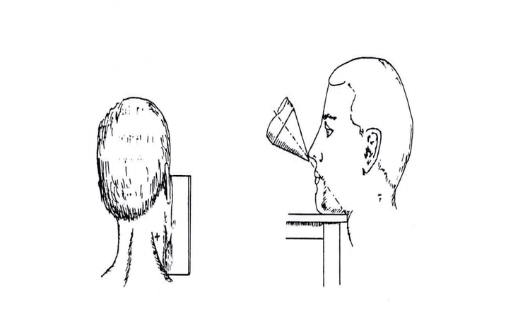
Adjust the shooting table, the ball is 0.5m from the film. The patient is instructed to lie on his stomach with his face pressed against the film by the edge of the eye socket, cheekbones, and nose on the side to be photographed. The frontal plane joins a 45 degree angle to the film, the Virchow plane is perpendicular to the film, the ear hole is in the middle of the film. The central ray is perpendicular to the major axis of the stony bone, going from the occipital bone to the cheekbone. The x-ray shadow is projected from the nape, the central ray lying in a plane perpendicular to the film. Stick the letter F on the corresponding digitized sensor plate to the right of the patient. Enter the patient's name, age, and gender into the machine, select the program that corresponds to the part to be taken. Adjust technical factors (70kV, 60mAs) Ask the patient to lie still for the scan. Close the door to the x-ray room. After taking the picture, instruct the patient to wait in the waiting room to get the results. Adjust the contrast, check the balance of the images on the film. Print film, compare with satisfactory film standards.
3. Evaluation of the results
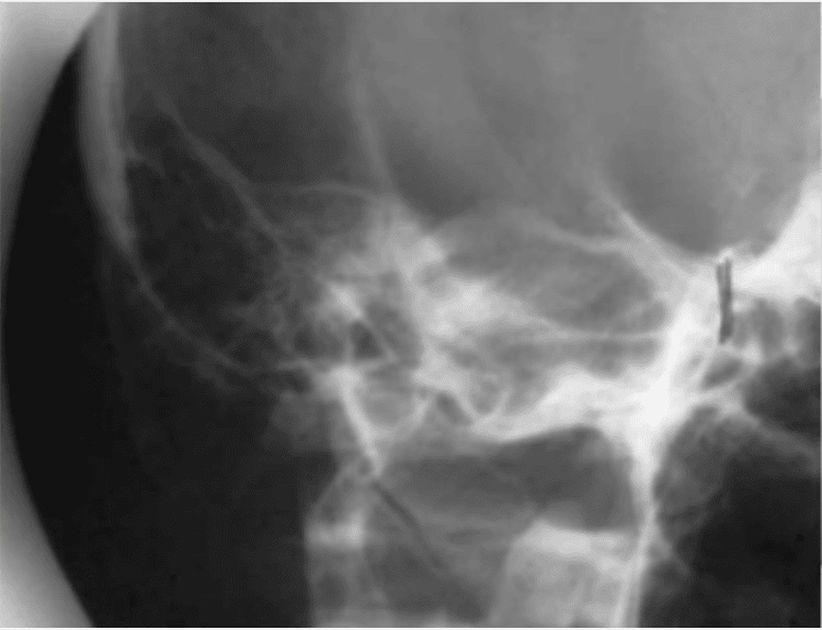
Standard film must clearly see the face of the stony bone, the lower 2/3 of the humerus and the posterior part of the occipital bone, and detect lesions if any. The doctor reads the lesion, describes it on a connected computer, prints the results and can provide professional advice to the patient and family if required. The technique of stenvers position x-ray does not cause complications for the patient. However, during the imaging process, the patient is not motionless, which will affect the quality of the film with unclear images.

Kết quả chụp xquang tư thế stenvers
Vinmec International General Hospital is one of the hospitals that not only ensures professional quality with a team of leading medical doctors, modern equipment and technology, but also stands out for its examination and consultation services. comprehensive and professional medical consultation and treatment; civilized, polite, safe and sterile medical examination and treatment space.
Customers who need advice on stenvers x-ray techniques, please contact Vinmec medical system here.


