This is an automatically translated article.
Article by Doctor Pham Quoc Thanh - Department of Diagnostic Imaging - Vinmec Hai Phong International General Hospital.
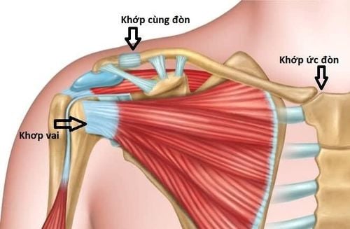
The sacroiliac joint is a movable joint formed by the distal clavicle and the apex of the shoulder, interspersed by a fibrocartilagen disc, covered anteriorly, posteriorly, above and below by a system of ligaments.
1. What is a joint strike?
Anterior and posterior ligaments are the strongest ligaments, reinforced by the fascia of the deltoid and trapezius muscles. The articular ligaments play an important role in stabilizing the anterior and posterior movements of the sacroiliac joint. The clavicle ligament consists of 2 ligaments, the ladder and the cone. These ligaments help limit the movement of the clavicle up and down.
2. Injuries to the sacroiliac joint are divided into several types
Sacral dislocation is a common shoulder injury (either from a sports injury or from a fall hitting the shoulder on hard ground). Common in cyclists, skiers or soccer players. A sacroiliac dislocation occurs when the force applied to the outside of the collarbone results in a mild, moderate or severe dislocation. In mild to moderate degree, the ligaments involved are stretched or partially ruptured. In contrast, in severe cases, the ligaments holding the clavicle down are broken, when the outer clavicle is pushed up, the outer skin can be seen.
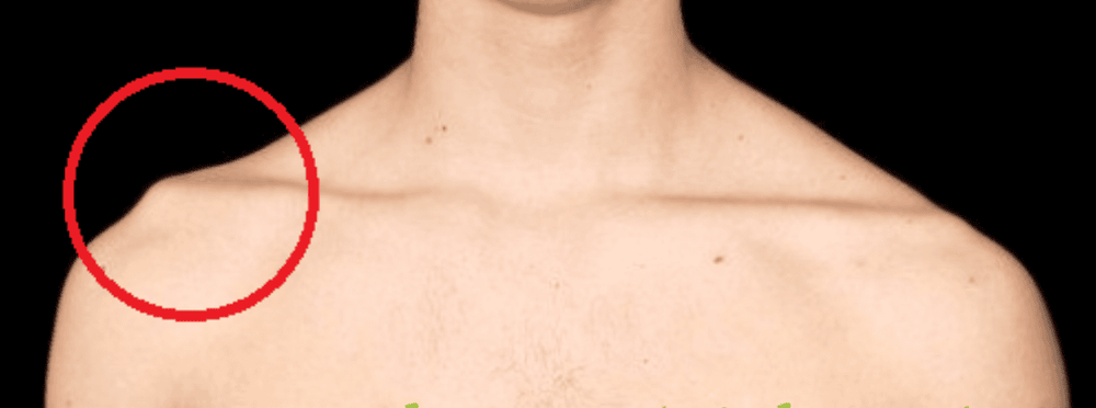
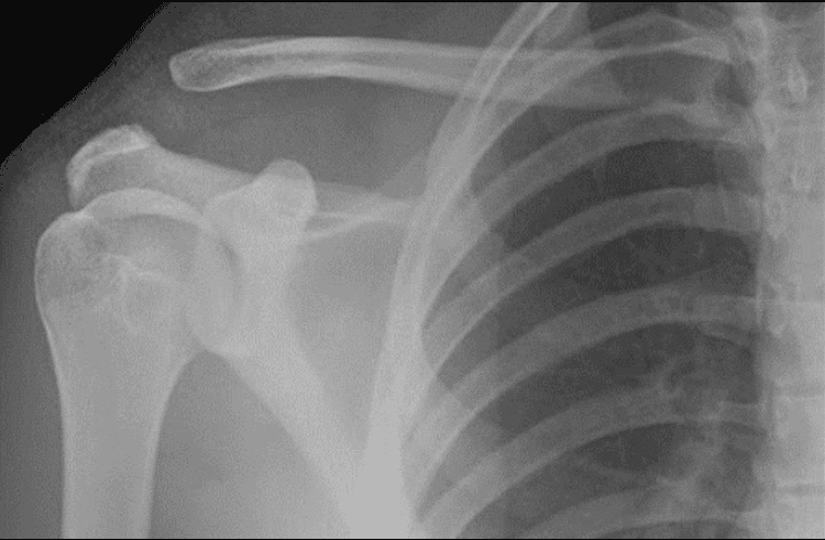
Classification
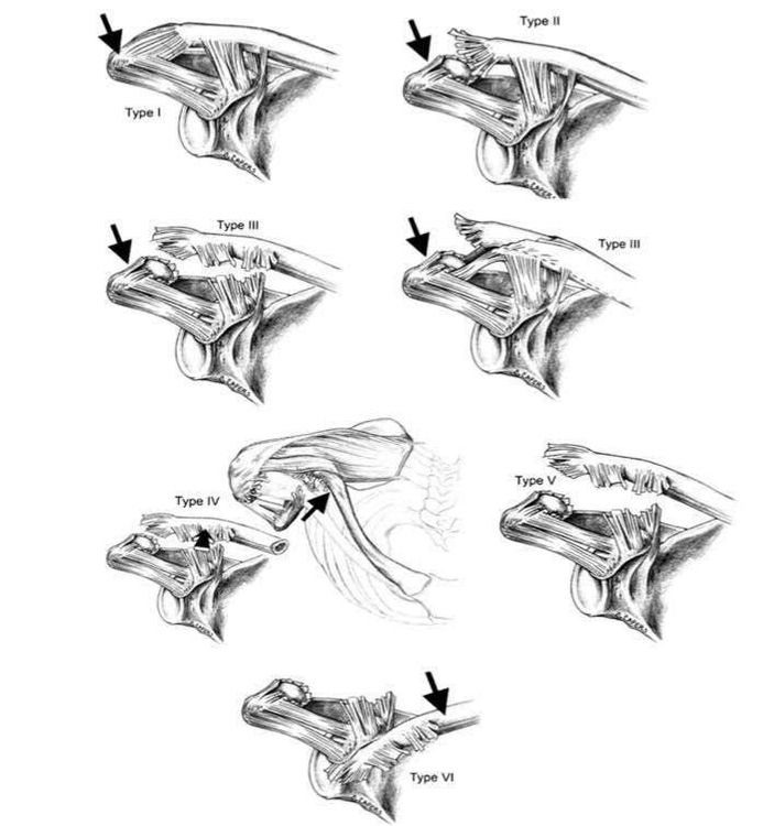
Tossy and Allman originally described the dislocations with 3 types I, II, III in 1960. Rockwood recorded and added to the classification of types IV,V,VI in 1984. according to the following table:
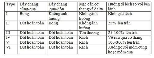
Accordingly:
Grade I is the stretching of the sacroclavicular ligament, the cochlear ligament is intact. Grade II: rupture of the sacroclavicular ligament, stretching of the cochlear ligament. Grade III: rupture of the sacroclavicular ligament, complete rupture of the clavicle ligament, grade III with the lateral clavicle displaced 25-100% relative to the contralateral side. Grade IV is that the lateral clavicle is displaced posteriorly into the trapezius muscle. Grade V is that the lateral clavicle is displaced by more than 100% of the contralateral side. Grade VI is rare with the lateral clavicle displaced into the inferior aspect of the crow's process.
3. X-ray image of sacroiliac dislocation in the anterior and posterior position
- Shoulder shooting with standard front-back, side-by-side poses, but Zanka pose gives the clearest view of the joint. The X-rays will be tilted about 10-15 degrees towards the head, to best observe the patient should carry about 5kg on each side and compare the two sides on the X-ray film.
High-intensity irradiation can allow the differentiation of lesions between type I and type II, which is especially important in distinguishing between type II and type III.
Image of dislocation with x-ray in front and back positions is a method that requires immediate examination to make an accurate diagnosis and provide the earliest treatment method.
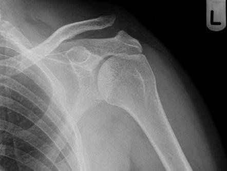
Modern X-ray machine system for clear images. The C-arm imaging system in the operating room cooperates with the surgeons to give the best image for the surgery to return the sacroiliac joint to its original position. Experienced KTVs, radiologists, together with skilled surgeons, best treat patients, a team of highly qualified, well-trained, specialized doctors at home and abroad. , experienced.
Please dial HOTLINE for more information or register for an appointment HERE. Download MyVinmec app to make appointments faster and to manage your bookings easily.


