This is an automatically translated article.
Bone cancer is one of the rare cancers. This type of disease is very dangerous and can be life-threatening because the time to detect the disease is often at a late stage.1. Overview of the skeletal system in the human body
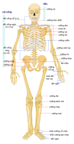
Normally, the human body has 206 bones of different lengths, which combine to form a skeleton to help support the body and protect internal organs from physical trauma. The skeletal system allows humans to move and function easily.
The human body has 3 main types of bones:
Long bones: tubular in shape, the middle will contain red marrow in children and yellow fat in adults. This is the most abundant bone in the human body, such as the femur, shin bone, and shin bone. Short bones: short in size, such as vertebrae, wrists, and ankles. Flat bones: semi-flat and thin, such as the skull and shoulder blades. Bones are connected to other bones by bands of tough tissue, called ligaments. Cartilage covers and protects the joints. Bones are hollow and filled with bone marrow.

Hệ thống xương trong cơ thể người
2. What is bone cancer?
Bone cancer is a rare cancer, often difficult to detect in its early stages, so the disease causes a high mortality rate.
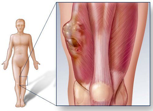
Bone cancer is a condition in which a malignant tumor appears in the bone. These cancer cells grow, compete with healthy bone tissue and can be life-threatening.
However, not all tumors are malignant. A person may have a benign tumor that, although it has not spread to other parts of the body, can grow large enough to press on surrounding tissues, weakening the bone and causing it to break.

Ung thư xương là tình trạng trong xương xuất hiện một khối u ác tính
3. Types of bone cancer
There are different types of bone cancer. Here are some common types of bone cancer:
Familial Ewing Sarcoma (ESFTs): this is the most common type of bone cancer. They mainly occur in children and young adults. Ewing sarcoma is usually present in bone, or soft tissues such as fat, muscle, fibrous tissue, blood vessels, or some other supporting tissue. This type of cancer usually occurs in the pelvis, along the spine, in the legs or arms. Cartilaginous sarcoma: is a type of cancer located in the cartilage tissue. Cartilage is a smooth, elastic tissue that covers and protects the ends of long bones in joints. Cartilaginous sarcomas mostly appear in the pelvis, shoulders, and thighs. Osteosarcoma: occurs in bone-like tissue (similar in structure to bone, but with less mineral content). This type of cancer usually occurs in the arms and knees.
4. Determine the stage of bone cancer
To determine the stage of bone cancer, doctors will use the TNM system:
Tumor (T): T is followed by numbers 0-4 used to describe size and location of the tumour. Tumor size is measured in cm.
Node (N): N stands for lymph nodes - small, bean-shaped organs that help fight infection. Lymph nodes near where the cancer started are called regional lymph nodes. Lymph nodes in other parts of the body are called distant lymph nodes. This stage is used to determine if the tumor has spread to the lymph nodes. If it has spread, where and how far has it spread?
NX: Regional lymph nodes cannot be evaluated. N0: Cancer has not spread to regional lymph nodes. N1: Cancer has spread to regional lymph nodes. This is rare for primary bone cancer. Metastasis (M): describes whether the cancer has spread to other parts of the body, called distant metastasis:
M0: The cancer has not spread. M1: The cancer has spread to another part of the body. M1a: Cancer has spread to the lung. M1b: Cancer has spread to other bones or organs.
5. Stages of bone cancer
Bone cancer includes the following 4 stages:
Stage I: The cancer is only in the bones and has not spread to other parts of the body. In this stage, cancer cells are less harmful and have not yet competed with normal cells. Stage II: The cancer cells have shown signs of growing stronger than before, but still limited to the bone. Stage III: More cancer cells (two to three sites) on the same bone tissue. Tumors at this stage can be highly or poorly differentiated. Stage IV: The cancer has spread from the bones to other places. These cells often grow very quickly and can affect other normal cells.
6. Diagnosis of bone cancer

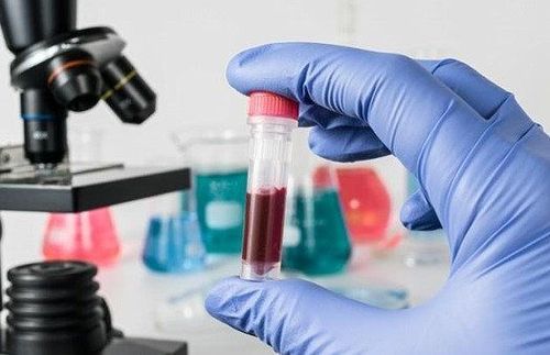
Xét nghiệm được coi là bước hết sức quan trọng nhằm xác định liệu bệnh nhân đã mắc ung thư hay chưa để sau đó đưa ra các chẩn đoán và tiên lượng bệnh
When a patient shows signs and symptoms of bone cancer, the doctor will make a diagnosis based on a physical exam and tests. The test is considered a very important step in determining whether a patient has cancer or not, and then making a diagnosis and prognosis. Here are some tests to diagnose bone cancer:
Blood tests: help find bone cancer. People with osteosarcoma or Ewing sarcoma may have higher levels of alkaline phosphatase and lactate dehydrogenase in their blood. However, it should be noted that high levels do not always mean cancer. Alkaline phosphatase is often high when the cells that form bone tissue are very active, such as when a child is growing or a broken bone is healing.
X-ray: is a way to create an image of structures inside the body using a small amount of radiation. It shows the original location of the cancer in the bone or where the cancer has grown in the body.
Bone scan: uses a radioactive tracer to look inside bones, helping to find bone cancer. The tracer is injected into the patient's vein. It collects in areas of bone and is detected by a specialized camera.
Computed tomography (CT or CAT) : takes pictures of the inside of the body using X-rays taken from different angles. A computer combines these images into a detailed 3-D image that shows the appearance of abnormal tumours. A CT scan can be used to measure tumor size.
Aspiration biopsy: take a sample of cells from the bone and look at it under a microscope to determine whether bone cancer is benign or malignant.
7. Treatment of bone cancer
Some of the main methods used in the treatment of bone cancer include:
Surgery: is the most common method in cancer treatment, can solve the root of the tumor, bring life to the person. sick. Radiation therapy: Using high-energy radiation to damage cancer cells and stop them from growing. Chemotherapy: uses drugs to kill dividing cancer cells. Bone scintigraphy is a method of early detection of lesions caused by primary bone cancer. From there, it is the basis for conducting biopsies and early detection of malignant tumors in the bone.
Nuclear Medicine Unit Room - Imaging Department of Vinmec Times City International Hospital provides Tc99m-MDP whole body bone scan service with many outstanding advantages compared to other imaging techniques other photo. Bone cancer, if detected early and properly treated, has a high chance of cure, reducing pain and costs for patients.
Customers can go directly to Vinmec Times City to visit or contact hotline 0243 9743 556 for support.
Articles refer to the source: cancer.net


