This is an automatically translated article.
Mammary gland disease is one of the diseases with high incidence in women, usually divided into two groups: Non-tumor mammary gland disease and tumor-induced mammary gland disease.1. Benign diseases of the mammary gland
1.1 Cystic fibrosis of the breast, also known as cystic fibrosis of the breast, one of the common benign lesions in women, is a localized tumor caused by fibrosis and hyperplasia of the mammary gland. Follicle formation occurs when there is an imbalance in the hormones estrogen, progesterone, and prolactin.
The fibrocystic breast tissue is flat, hard, and mobile; feel soft masses, unclear boundaries appear in the second half of the menstrual period, in the outer half of the breast or maybe both breasts. These changes cause the breasts to become thickened, painful, and tight.
Nearly half of women with cystic fibrosis have no symptoms. Some common symptoms include cyclical pain and swelling of the breasts. When squeezing, the breast feels full, stagnate, painful, and tight. The breast tissue is dense and feels lumpy like pebbles. Symptoms of breast tenderness, increased breast size, and multiple cystic fibrosis must be distinguished from benign fibrocystic masses or lesions of breast cancer.
1.2 Fibroids of the breast is a common benign disease in young women in the first 20 years after puberty, many cysts can occur in one or both breasts.
Clinically a typical fibrocystic breast is usually a round, firm, firm, well-demarcated mass, movable, painless, varying in size from 1-5 cm.
The development of fibrocystic breast is quite easy and is often spontaneously discovered, but in women over 30 years of age, it should be distinguished from breast cysts or breast cancer.
1.3 Cystosarcoma phyllodes Is a type of fibrocystic breast with stromal cells that grow very quickly to form a very large, hard tumor, even occupying the entire breast. The disease is usually benign, but there is a small incidence of malignancy.
Treatment is usually surgical removal of the entire tumor and the surrounding healthy tissue to prevent recurrence.
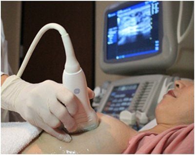
1.4 Breast cyst is a disease often related to endocrine. Cysts are discrete, well-defined, mobile, dense masses. The size of the cyst is different, often there are many cysts in one or both breasts, when ultrasound will show homogenous hypoechoic areas, clearly demarcated, thin shell.

Bệnh nang vú liên quan đến nội tiết
1.5 Fat tissue necrosis is a rare disease that usually occurs after trauma or biopsy. Pain may or may not be present. The tumor may go away on its own without treatment.
1.6 Breast Abscess The phenomenon of breast abscess is the appearance of a red, painful, non-hardening area due to the back infection of the mammary ducts and then spreading around.
Because breast abscess often occurs during pregnancy or lactation, the early stages of inflammation can still be breastfed and at the same time treated with antibiotics as prescribed by the doctor.
If it comes to a late stage, the inflammatory mass can turn localized pus into a painful and concave mass, sometimes it can burst the pus mass on its own. In this case, it is necessary to open the pus mass and drain it.
1.7 Nipple discharge Most cases of abnormal breast discharge are associated with ductal papilloma, ductal cystic fibrosis, or breast cancer.
1.8 Breast deformity Unilateral or bilateral breast enlargement often requires plastic surgery to reduce it.
2. Malignant breast disease and what you need to know
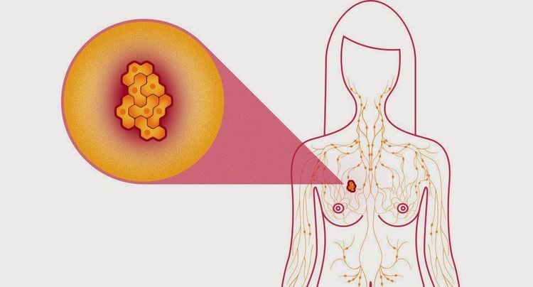
Malignant breast tumor (mammary gland cancer) is an abnormal change in the cells of breast tissue, they initially arise in one location, then can invade and destroy nearby tissue and organs. . There are two main types of breast cancer: ductal cancer and lobular cancer.
70% of breast cancer cases detected: Single, non-tense, firm, with indefinable margins. Besides, 90% of tumors are detected by the patients themselves, especially small tumors < 1cm.
Less common symptoms are breast pain, discharge. Late detection, especially when the tumor is larger than 5cm, can see the tumor shrinking the skin and nipple, breast enlargement, orange-like skin, pain, tumor sticking to the skin or chest wall, examining the axillary pit. bubonic. There are even ulcers, supraclavicular lymph nodes, hand edema, bone metastases, lung, liver, brain...
2.1 Subclinical diagnosis Mammography: It can be detected very early from 2 years before palpation. About 35% of cases are detected by screening mammography alone. Breast MRI helps to determine the tumor's nature, the border is not clear with the surrounding tissues, there is an increase in angiogenesis and the degree of invasion of the tumor to the surrounding, especially compared to the chest muscle. Biopsy: The gold standard for cancer identification and classification because 30% think of cancer clinically but biopsies are benign and 15% think benign but biopsies are malignant. Technetium 99m bone scintigraphy - labeled phosphonates, is an important means of evaluating breast metastases. The rate of bone metastases increases with disease stage, if Stages I and II are only about 7% and 8% have (+), while stage III is 25% (+). 2.2 Special Types of Breast Cancer 2.2.1 Paget's Disease There is a change in skin color in the nipple area in the form of eczema and 99% of it is malignant. The underlying tumor was palpable, 95% of which were found to be due to metastatic cancer, most of which were due to infiltrating ductal carcinoma.
Paget's disease is rare but often detected late because the symptoms are not clear. Treatment can include mastectomy and postoperative radiation.
2.2.2 Inflammatory breast cancer is found as a lesion in the skin of the breast, redness in the surrounding margin, and usually no palpable tumor below.
This is a malignant disease because when detected, up to 35% of cases have metastases, but fortunately, the disease only accounts for < 5% of breast cancers.

Ung thư vú
2.2.3 Breast cancer during pregnancy and lactation In most cases, mastectomy is the minimal treatment modality of choice. If the pregnancy is later than in the last 3 months of pregnancy, it is possible to remove the tumor and radiation immediately after birth.
2.2.4 Bilateral breast cancer Rate is only about <1%, but there is a higher rate of 5-8% for stage 2 breast cancer cases. Bilateral breast cancer is common in women <1% 50 years old and usually lobular cancer.
2.3 Measures for early detection of breast cancer Clinical examination and mammography detect up to 40% of early-stage cancers and another 40% by palpation. Mammograms may be done every two to three years in women aged 40-49 years and annually in older women.
Breast ultrasound usually only helps in the differential diagnosis of cysts and fibroids. Ultrasound should only be considered as an adjunct to clinical examination and mammography in breast cancer screening. Genetic testing: In patients with a family history of breast cancer, the positive detection of 2 genes BRCA1 and BRCA2 is associated with an increased risk of breast cancer as well as ovarian, colon, and prostate cancers. prostate and pancreatic cancer. 2.4 Treatment of breast cancer Extensive mastectomy is often indicated in the early stages of the disease to remove the tumor or part of the breast or a quarter of the breast to save.
Conservative treatment if there is only one lesion <2 cm in diameter. However, conservative treatment must be accompanied by immediate histological examination of the surrounding margin to ensure complete resection of the cancerous tissue. Patey mastectomy is often indicated in Vietnam, along with axillary lymph node dissection. This method is indicated regardless of the stage of the disease. Radiation, chemotherapy, and endocrine therapy are adjuvant therapies in breast cancer. To register for examination and treatment at Vinmec International General Hospital, you can contact Vinmec Health System nationwide, or register online HERE.
SEE ALSO :
Treatment of cystic fibrosis of the breast Causes and signs of cystic fibrosis of the breast Instructions for periodic breast self-examination at home


