This is an automatically translated article.
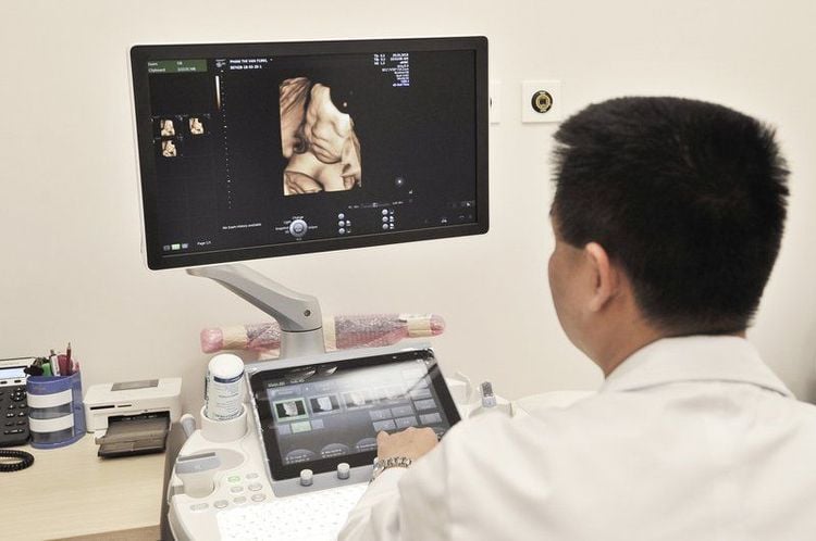
Written by Master, Doctor Nguyen Ngoc Tu, Doctor of Obstetrics and Gynecology, Department of Fetal Medicine, Center for Obstetrics and Gynecology - Vinmec Times City International General HospitalFetal ultrasound uses ultrasound waves to image and observe the structure and activity of the fetus in the uterus. During the ultrasound, the doctor will use an ultrasound probe placed on the pregnant woman's abdomen (trans-abdominal ultrasound) or use an ultrasound probe covered by a condom to insert into the pregnant woman's vagina (first line ultrasound). transvaginal probe) to conduct an ultrasound.
1. How is a fetal ultrasound performed?
Ultrasound is performed in a low-light room to ensure the best image displayed on the machine. You will be lying on the ultrasound bed, exposing the abdomen where the ultrasound is needed (trans-abdominal ultrasound) or exposing the private area (transvaginal ultrasound)
The doctor will apply a little gel on the area to be ultrasound sound to ensure the best sound wave transmission. The image of the fetus displayed on the screen in 2 colors black and white

Siêu âm thai không gây đau đớn và giúp mẹ bầu thấy được hình ảnh thai nhi
2. Pregnant women need to prepare before ultrasound?
Ultrasound is best done when the bladder is not full (except in cases of transabdominal ultrasound in early pregnancy). Therefore, it is necessary to urinate before the ultrasound. Do not apply body lotion/oil for 48 hours before the ultrasound.
Ultrasound is painless. However, in some cases, the doctor presses the transducer slightly, which can cause discomfort.
Details of fetal development week by week, every parent should learn:
Trắc nghiệm: Bạn có biết nên khám thai lần đầu vào lúc nào không?
Việc khám thai lần đầu mang ý nghĩa rất quan trọng, giúp bạn xác định chính xác mình có mang thai hay không? Thai nhi đã vào buồng tử cung hay chưa?... Vì vậy, nếu chưa biết khám thai lần đầu vào lúc nào, trả lời nhanh 5 câu hỏi trắc nghiệm sau sẽ giúp bạn có câu trả lời.3. Is fetal ultrasound safe?
Ultrasound has been used in obstetrics and gynecology for decades and is considered safe during pregnancy, and has a full set of standard and procedure guidelines. Follow that standard, so far, no one has recorded ultrasound harm to the fetus
No effect on miscarriage or effect on the fetus when using a vaginal transducer, even with bleeding vagina.
4. When is fetal ultrasound done?
Ultrasound can be done at any time during pregnancy. Depending on each specific case, the doctor can specify the time and number of ultrasounds for each person differently, there are 3 times during pregnancy that experts recommend really necessary:
From 12 weeks up to 13 weeks 6 days From 19 weeks to 22 weeks From 30 weeks to 32 weeks
5. How long does a pregnancy ultrasound last?
Ultrasound screening for fetal malformations usually takes about 15-30 minutes. In some cases when the fetus is difficult to assess due to difficult posture or too much movement, or the thick layer of abdominal wall tissue obstructs, making it more difficult for ultrasound waves to pass through, so the ultrasound time may have to be longer. or make an appointment for the next time.
6. Why do pregnant women need ultrasound in early pregnancy?
Assess for intrauterine or ectopic pregnancy Assess for fetal heart rate Assess number of fetuses Expected delivery Evaluate uterine and adnexal abnormalities Ultrasound assess cause of vaginal bleeding
7. What does an ultrasound at 12 weeks to 13 weeks 6 days include?
Assess gestational age and expected birth according to head and buttocks length (which is the most accurate assessment time).
Screening for chromosomal abnormalities:
Measure the nuchal translucency (NT). The nuchal translucency is a layer of fluid under the skin at the back of the baby's nape. All fetuses have this fluid, but many fetuses with chromosomal abnormalities show signs of increased thickness of this fluid. The NT measurement combined with the Double test blood test can screen for about 90% of fetal Down syndrome cases. Early observation of anatomical structures such as arms, legs, abdominal wall, heart, skull, placenta... to detect major abnormalities of the fetus
Evaluation of opacity in the brain (IT) to detect early neural tube defects.
Screening for preeclampsia
8. What does a 19-week to 22-week fetal ultrasound include?
Is the best time in pregnancy to evaluate structural abnormalities of the fetus. 19 weeks is the time when a detailed assessment of fetal structure can be started and 22 weeks is the best time for evaluation. Observe the morphology and structure of the skull and brain, face, digestive system like liver, intestines, respiratory system like lungs, diaphragm, circulatory system like heart, urinary system like kidneys, bladder, etc. skeletal system and limbs, sex... Observe the placenta, umbilical cord and amniotic fluid Measure the length of the cervix to assess the risk of premature birth Assess the baby's development status by measuring biological indicators of the baby such as biparietal diameter, head circumference, femur length, abdominal girth... to assess whether the baby's development corresponds to gestational age, is it small and if it is small, what are the risks? or not.

Siêu âm thai đánh giá được tình trạng sức khỏe cũng như các dị tật của thai nhi
9. What does a 30 to 32 week pregnancy ultrasound include?
It is an important time to assess fetal growth.
Like at 22 weeks, the doctor will measure the biological indicators of the fetus to assess whether the fetus is developing normally, or if the fetus is small or larger than normal
Fetal weight will be shown in the newspaper. Report on the percentile line
The 10th percentile line is for a normal weight baby. The lower 10th percentile line is a small baby. The 90th percentile line is a big baby. The weight on the ultrasound has deviations from the real thing. about 10%
Use Doppler ultrasound to assess fetal circulation through major arteries. From there, assess the risk of hypoxia, impaired placental function of the fetus
With a small fetus, there may be a bad prognosis for the fetus. To be able to have a sure diagnosis as well as give the best management, the doctor may order additional ultrasound and monitor the fetal growth rate through ultrasounds.
With large fetuses, usually those older than gestational age are born healthy. However, a small number of large pregnancies are caused by maternal metabolic disorders such as gestational diabetes, which, if not detected, can cause dangerous complications for both mother and child. To be safe during pregnancy, the doctor will order more necessary tests.
Assessment of fetal organ structures is the same as at 22 weeks and adds some differences such as assessment of late-onset lesions such as cortical lesions, intestinal obstruction, ureteral pyelonephritis, infection Zika, CMV, Rubella...
10. Can ultrasound detect all abnormalities of the fetus?
Depending on the disease, there are diseases that are easier to detect, there are diseases that depend on the level of expression, the progression changes, it is difficult to detect even undetectable on ultrasound.
Some abnormalities such as heart abnormalities or intestinal obstruction... can often only be detected in the late stages of pregnancy. Ultrasound screening for fetal malformations, although it can rule out most of the above-mentioned abnormalities, cannot rule out all fetal abnormalities. Diseases such as autism, cerebral palsy, mental retardation cannot be detected on ultrasound because these diseases are not caused by structural abnormalities.
11. If the ultrasound results have problems, what should the pregnant woman do?
It's only natural to be worried when your ultrasound shows a problem with your pregnancy. However, problems on sonography can range from large, obvious, serious abnormalities to minor problems, which are just structural variations with no consequences. So when ultrasound suspects a problem, you will be transferred to a specialist in fetal medicine for advice and guidance on diagnosis and treatment in detail.
Fetal abnormalities, which can be treated, may not, some can be treated right from the fetal stage, some have to wait until birth to be treated. Depending on the abnormality, you will be guided and consulted step by step: what to pay attention to do next in pregnancy, make a birth plan for you, make a plan for postpartum care for your baby, make a plan Surgical treatment if necessary, instructions for taking care of the baby after discharge... Regardless of the type, this is also the time when you need the most support, not only medical. The first 3 months are the most sensitive time during pregnancy. In order for mother and baby to be healthy, parents need to pay attention to:
Understand early signs of pregnancy, pregnancy poisoning, bleeding during pregnancy. Timely, correct and sufficient first prenatal check-up, avoiding too early/too late. Fetal malformation screening at 12 weeks detects dangerous fetal malformations that can be intervened early. Distinguish between normal vaginal bleeding and pathological vaginal bleeding for timely intervention to maintain pregnancy. Screening for thyroid disease in the first 3 months of pregnancy avoids dangerous risks before and during delivery. Vinmec currently has many maternity packages (12-27-36 weeks), in which the 12-week maternity package helps monitor the health of mother and baby right from the beginning of pregnancy, early detection and timely intervention of health issues. In addition to the usual services, the maternity monitoring program from 12 weeks has special services that other maternity packages do not have such as: Double Test or Triple Test to screen for fetal malformations; Quantitative angiogenesis factor test for preeclampsia; thyroid screening test; Rubella test; Testing for parasites transmitted from mother to child seriously affects the baby's brain and physical development after birth.
Please dial HOTLINE for more information or register for an appointment HERE. Download MyVinmec app to make appointments faster and to manage your bookings easily.


