This is an automatically translated article.
Eyeball adhesions is a condition in which fibrous formations adhere to the conjunctiva of the eyeball to the ciliary conjunctiva, making both the eyelids and the eyeball unable to move, which can be caused by burns, tumors, malformations, .. To treat blepharospasm, surgery is required. Read more articles below about eyelid surgery method.
1. What is eyelash extensions?
The phenomenon of eyeball adhesion causes the eye to deviate and limit eye movement. The consequence if this condition goes on for a long time is the loss of vision function. Moreover, if the eye condition is very deviated and can't see everything for a long time, it will cause amblyopia. Therefore, it is necessary to relieve the eyeball adhesions surgically.
Time to carry out surgery again to separate the eyeball adhesions must be at least 6 months apart from the previous tumor surgery. If possible, it may take longer for the tissues to completely heal. There are many causes of this condition such as eye burns, tumors forming in the eye or surrounding area, deformities,... In which, eye burns are a special emergency in ophthalmology, especially chemical burns, because in many cases, even with prompt and aggressive treatment, blindness cannot be prevented. Treatment of eye burns must be treated urgently and quickly to preserve the physiological function of the eye. The prognosis of a burned eye, whether good or bad, depends very much on the initial emergency management. Severe or mild eye damage depends on the time the chemical stays in the eye, the concentration of the chemical, the physicochemical properties of the chemical that changes the pH of the eye, and destroys proteins.
In case of minor burns: Irritated eyes, conjunctival swelling, corneal epithelium edema, visual acuity decreased slightly.
For severe burns: The eyes are painful, the conjunctiva is edematous, occluded, and there are white necrotic places without blood vessels. The cornea is opaque white, the posterior part cannot be seen. The corneal epithelium is shed, the tissue is opaque and edematous, the corneal ulcer is persistent and untreated, leading to eye perforation, vision is drastically reduced, and only dim light can be distinguished.

Hiện tượng dính mi cầu làm mắt lệch đi và hạn chế vận động của mắt, ảnh hưởng đến chức năng thị giác
2. Eyelid detachment surgery
Eyelid detachment surgery is indicated for patients with the following characteristics:
Severe blepharospasm affecting eye movement and vision problems Sequelae of eye burns, trachoma. Stevens-Johnson syndrome, Pemphigoid. Recurrence of blepharospasm after ocular surface surgery. Some cases are not indicated for blepharoplasty surgery include:
Patients with severe eye infections such as: acute infectious keratoconjunctivitis, necrosis, need anti-infective treatment. In patients with severe deformity or deficiency of the eyelid margin that causes blepharoplasty, which will lead to surgical failure, blepharoplasty should be performed prior to blepharoplasty. Patients with systemic diseases also need to be considered when performing surgery because of the influence of the underlying disease on the process before, during and after surgery. 2.1. Steps Performer The person performing the surgery is an Ophthalmologist who has been trained in this surgery
Equipment Instruments include: A set of microsurgery instruments, automatic eyelid rims, large needle forceps, rectoid fixation needle, conjunctival dissection forceps, hemostatic forceps, bipolar electrocautery, 8-0, 9-0, 10-0 sutures, blood-absorbing gelaspon, plastic mold or contact lenses if needed.
Drugs used in surgery: Local anesthetic (surfactant or injectable adjacent to the eyeball), eyewash solution (physiological salt or ringer lactate)
Patient For the patient, the doctor will check the muscle condition. body prior to surgery. Indicated to drink and apply medication before surgery. After that, the patient is dressed in surgical clothes, and the eye area and eyelids are cleaned.
2.2. Steps to carry out surgery Check the records as prescribed. Check the patient: Check the patient's condition such as blood pressure, heart rate, ... and especially check the eye condition before surgery. 2.3. Performing the Anesthesia technique is also known as the anesthetic step. For children, the doctor will administer general anesthesia, while for adults, local anesthesia will be given with topical anesthetic and paraocular injection (Lidocaine 2% or xylocaine 2%). In severe cases with prolonged or poorly cooperated prognosis by doctors, pre-anesthesia or anesthesia may be used.
Techniques used: Step 1: Separation of the blepharospasm, exposing the rectus muscles in the area with fibrous adhesions if necessary, conducting dissection and resection of the subconjunctival fibrous tissue, burning to stop bleeding. Apply anti-metabolites as indicated to the fibrous adhesions for 3 minutes, rinse with ringer lactate solution.
Step 2: Carry out conjunctival dissection of the healthy conjunctiva, the size of the graft is equivalent to the size of the area to be grafted
Step 3: Move the graft to the area to be patched, doctors need to avoid flipping the graft. Then proceed to graft into the sclera with 8-0 or 9-0 or 10-0 loose stitches.
Step 4: At the end of surgery, the doctors check the adhesion of the conjunctiva, the width of the corners and the furniture, place a non-stick plastic mold or contact lenses if necessary. Nurses can help patients administer antibiotics and change the compression bandage.
2.4. Treatment and monitoring After surgery, the patient was prescribed medication by the doctor, including antibiotics, corneal nutrition, and anti-inflammatory corticosteroids. The doctor needs to monitor the adhesion of the conjunctiva, the width of the corners of the map, the epithelialization of the surface of the eyeball.
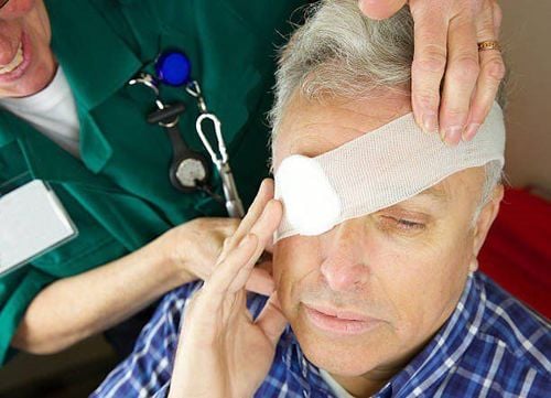
Sau khi phẫu thuật tách dính mi cầu, bệnh nhân sẽ được bác sĩ kê đơn thuốc và theo dõi tình trạng
2.5. Complications that can occur during and after surgery During surgery Bleeding can occur if the doctor touches the rectus muscle by pressing on the bleeding area, if this does not stop the bleeding can be stopped. .
Perforation of sclera or cornea during deep dissection: Restoration with 9-0 or 10-0 sutures, can use multi-layer amniotic membrane or corneal and scleral transplant if grafting material is available.
After surgery If the patient has edema of eyelids, conjunctiva and graft, use hypertonic solution and anti-edema drug.
When the patient has a hematoma, bleeding under the graft, it is necessary to take hemolytic drugs, hemostatic drugs such as ventricular, vitamin C, transamin, adrenoxyl,... If the hematoma persists for more than 5 days after the graft, Surgery can remove the hematoma.
Loss of thread, detachment of graft: For partial detachment, contact lenses can be placed and monitored, if the condition is large, stitches should be fixed to fix the graft
To examine and perform de-adhesion surgery eyelids, you can go to the eye specialist - Vinmec International General Hospital. The department has a comprehensive vision and eye health care function for children, adults and the elderly including refractive error testing, general examination, diagnostic ultrasound, laser treatment and surgery. In addition, ophthalmology also has the task of coordinating with other clinical departments in the treatment of pathological complications and eye injuries caused by accidents.
Why should you choose to examine and treat eye diseases at Vinmec International General Hospital?
Simple and quick procedure. Enthusiastic advice and support, reasonable and convenient examination process. Comprehensive facilities, including a system of clinics and consultations, blood collection room, dining room, waiting area for customers... The medical staff has high professional qualifications, style. Professional, caring way of working.
Please dial HOTLINE for more information or register for an appointment HERE. Download MyVinmec app to make appointments faster and to manage your bookings easily.


