This is an automatically translated article.
Posted by Doctor Ta Que Phuong - Department of General Internal Medicine - Vinmec Times City International HospitalThe gastrointestinal foreign body is an endoscopic gastrointestinal emergency. This accident is common in children under 5 years old. There are many different causes of digestive problems in children. Digestive foreign bodies are often caused by children not actively swallowing but by accidently swallowing objects that are too large or angular, falling into the digestive tract and cannot be digested.
1. When is urgent removal indicated?
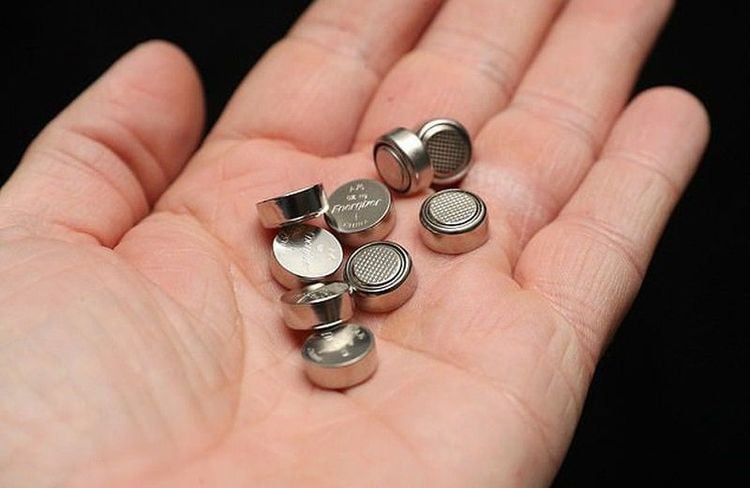
Urgent intervention (ie, endoscopic or other surgical removal of the foreign body) is indicated if any of the following warning signs are present:When the patient presents with signs of airway risk When there is evidence of near-complete esophageal obstruction (eg, patient is unable to swallow secretions) When a sharp, long (>5 cm) or superabsorbent polymer is lodged in the esophagus or stomach When the foreign body is ingested input is a magnet or a high-powered magnet When the disc battery is in the esophagus in some cases in the stomach) When signs or symptoms suggestive of inflammation or bowel obstruction (fever, abdominal pain or vomiting)

2. How to evaluate gastrointestinal foreign bodies?
A careful clinical examination is key in the diagnosis of an esophageal foreign body and for the prevention of its complications. If symptoms are present, they may suggest the likely location of the foreign body. Imaging is used to detect and locate foreign bodies. The diagnostic and therapeutic steps depend on the patient's symptoms, the shape and location of the foreign body3. Clinical examination
The airway and breathing rate should always be checked first. Examination of the neck may reveal swelling, erythema, or crepitus, which suggests that esophageal perforation has occurred, in which case surgical consultation is required. Examination of the chest may reveal signs of breathing or wheezing, suggesting an esophageal foreign body with tracheal compression. Abdominal examination may reveal evidence of small bowel obstruction or perforation, in which case immediate surgical consultation and abdominal imaging are required.A hand-held metal detector has been used with varying success in locating coins, but this is rarely used in clinical practice because coins are easily detected by radiography usually, common, normal. Metal detectors can be useful for detecting materials that are metallic but not radiopaque, such as aluminum, but this use is limited because the instrument is less reliable at detecting objects. metals other than coins.

4. Diagnostic Imaging
Conventional radiographs For all patients with suspected foreign body ingestion, the initial diagnostic test should be biplane radiographs (anterior and posterior) of the neck, chest, and abdomen. It is recommended to include each of these sites even if symptoms suggest a site (eg, esophageal or respiratory symptoms). Likewise, it is recommended that routine radiographs be performed even if the foreign body is thought to be radioactive. This is to assess the likelihood of other swallowed objects, for indirect evidence of radioactive foreign bodies (such as air-fluid levels in the esophagus), and for free air to represent perforation. Toys made of plastic or wood, some thin metal objects, and many types of bones that are not easily seen on a regular X-ray machine. In a study in children, only 64 % of ingested foreign bodies were radiopaque. Other foreign bodies, such as "fidget spinner" toys, which have both radioactive and radioactive components, may break apart and progress separately in the gastrointestinal tract.CT scanner or MRI If plain X-ray does not reveal any foreign body or abnormalities, further evaluation depends on patient characteristics and suspected foreign body:
Computed tomography (CT) or scan magnetic resonance imaging (MRI) - If the patient is symptomatic, or if a foreign body is suspected, any dangerous features are large > 2 cm wide, > 5 cm long, or sharp), or if the type of anomaly objects not clearly known to the caregiver, CT with three-dimensional reconstruction should be used as the follow-up diagnostic tool. Alternatively, MRI can be used to evaluate objects that are radioactive but is contraindicated if any metallic foreign bodies are present.
There is no need for CT or MRI imaging if the patient is completely asymptomatic, the foreign body has small benign features <2 cm, is not sharp or elongated and is not a magnet or pin), and the emitter now certain of the type of foreign body ingested. In this case, it is reasonable to discharge the patient after a period of healthcare observation, if the patient remains completely asymptomatic and is able to eat and drink normally.
Vinmec International General Hospital is a high-quality medical facility in Vietnam with a team of highly qualified medical professionals, well-trained, domestic and foreign, and experienced.
A system of modern and advanced medical equipment, possessing many of the best machines in the world, helping to detect many difficult and dangerous diseases in a short time, supporting the diagnosis and treatment of doctors the most effective. The hospital space is designed according to 5-star hotel standards, giving patients comfort, friendliness and peace of mind.
Please dial HOTLINE for more information or register for an appointment HERE. Download MyVinmec app to make appointments faster and to manage your bookings easily.


