This is an automatically translated article.
Posted by Doctor Mai Vien Phuong - Department of Examination & Internal Medicine - Vinmec Central Park International Hospital
White blood cells circulate in the blood circulation and help the immune system fight infections. When an infection or inflammation occurs, the body releases white blood cells to help fight the infection.
1. What are eosinophils?
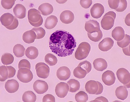
Eosinophils are one of the three components of granulocytes, which are produced in the bone marrow and are one of the cells that play a role in promoting inflammation, especially allergic inflammatory reactions. Therefore, large quantities of eosin can accumulate in tissues such as the esophagus, stomach, small intestine and sometimes in the blood when such individuals are exposed to the allergen. The allergens that cause this EE esophagitis are not known, only that they enter through inhalation or ingestion.
Although eosinophils are part of the immune system, their response is not always good for the body. Sometimes, they are part of conditions such as food allergies, inflammation in the body's tissues.
2. Where is the stomach and intestines located?
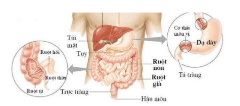
The stomach, also known as the stomach, is a part of the human digestive system. This organ is shaped like a bag, similar to the letter "J" and is the largest bulge of the digestive system. The stomach is located in the upper left part of the abdominal cavity, just below the liver and next to the spleen. In the digestive system, the stomach is located between the esophagus and the duodenum. This is where food comes from the esophagus. After food enters the stomach, it secretes acids and enzymes to digest the food. Besides, the muscles of the stomach will periodically contract, turning the food upside down to enhance digestion.Intestine is a general term, referring to the small intestine, large intestine (colon) and rectum
The small intestine is a part of the digestive tract and of the digestive system. In addition to the small intestine, the alimentary canal also includes the large intestine, stomach, mouth, pharynx, rectum, anal canal, and anus. In the human body, the small intestine is located behind the stomach and before the large intestine (also known as the colon)
The colon, also known as the large intestine, is the near-terminal part of the digestive system, attached to the last part of the digestive system. Chemistry is the anal canal, which is an extremely important part of the abdomen.
Winding into a frame. The colonic framework consists of the right colon starting from the cecum where the small intestine empties into the (very short) large intestine. It joins the transverse colon and descends to the left colon, and finally to the sigmoid colon (very short) to the rectum.
3. What is gastroenteritis?
Gastroenteritis is an inflammatory lesion of the inner lining of the stomach and small intestine and colon. Most cases are caused by infection, although gastroenteritis can occur following ingestion of drugs and ingestion of toxic chemicals (eg, metals, pathogens). Gastrointestinal tract infection, gastroenteritis can cause diarrhea from mild to severe, serious illness when not treated promptly.
Viruses are the leading cause of gastroenteritis, in the UK 2 strains of viruses cause this disease: Norovirus and Adenvirus. The virus is present after we go to the toilet, easily spread from person to person by direct contact or indirect contact through touching objects.
Food poisoning from eating food contaminated with viruses can also cause gastroenteritis. Common pathogens are Campylobacter, Salmonella and E.Coli. Toxins secreted by bacteria can also cause poisoning, some groups of parasitic organisms can also be the cause of the disease.
Recently, however, scientists have found a new form of the disease, which is eosinophilic gastroenteritis
4. What is eosinophilic gastroenteritis?
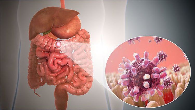
Eosinophilic gastroenteritis (VTQDBCAT) is an eosinophilic infiltrate in the stomach, small intestine, colon, and rectum causing clinical symptoms.
Eosinophilic gastroenteritis is a rare and unusual condition characterized by patchy or diffuse eosinophilic infiltration of the gastrointestinal tract, which was first described. described by author Kaijser in 1937. Each case is unique with specific lesions, depending on the location as well as the depth and depth, extensive spread of the intestine and chronic recurrence (chronic) relapsing course ). The disease can be classified into mucosal, muscular, and serosal types based on the depth of the lesion. Reports are adequate and damage to almost all digestive organs from esophagus to colon, in which stomach is the most affected organ, followed by small intestine and colon.
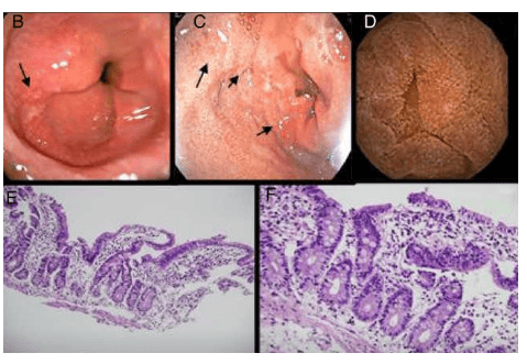
Eosinophilic gastroenteritis (English used from Eosinophilic gastroenteritis_EG) is a disease in the following group of diseases: eosinophilic esophagitis (eosinophilic esophagitis), gastritis, enteritis, colitis eosinophil gastritis, enteritis, and colitis), and members of this family of diseases are collectively known as eosinophilic gastrointestinal disorders (EGIDs). Although rare, eosinophilic gastroenteritis should be recognized by the clinician as soon as possible because the disease presents with signs, symptoms, and syndromes that are "similar" to some other diseases. other digestive disorders, especially peptic ulcers, but with proper treatment, the disease is still not cured. Furthermore, eosinophilic gastroenteritis is treatable, sometimes presenting as masquerade as the irritable bowel syndrome. The diagnosis of EG is confirmed by biopsy or collection of eosinophilic ascites in the absence of other pathogens, including intestinal parasites. Eosinophilic esophagitis should also be differentially diagnosed and discussed. It is also important to note that the disease often manifests itself in one or more members of a patient's family.
5. How is eosinophilic gastroenteritis found?
The concept of VDDRDBCAT was first described in 1937 by the author Kaijser, but it was not until 1961 that Ureles et al. first classified this pathology into two types: diffuse and localized, forming tumors seed. In 1970, KLein et al. divided this pathology into three types based on the degree of infiltration of BCAT: mucosal, myosin, and serosa. By 1985, Oyaizu et al. had found evidence to confirm the role of IgE, mast cell-mediated mechanisms in the pathogenesis of VDDRDBCAT. Further, case series studies and longitudinal follow-up studies have contributed to further knowledge of the clinical symptoms, histopathology, treatment, and course of this rare disease. By definition, it is a disease characterized by chronic inflammation of the gastrointestinal tract due to eosinophilic infiltration, which may occur in the stomach, small intestine, colon, or rectum. Peripheral blood eosinophil counts may or may not be increased.
Source: http://www.revistagastroenterologiamexico.org/en-eosinophilic-gastroenteritis-an-unusual-presentation-articulo
6. Is eosinophilic gastroenteritis common?
Epidemiologically, this is a disease with a relatively low rate ranging from 1-20 cases/100,000 people. The disease occurs in both children and adults, but it is most common in the age group 30 - 50. The male/female distribution ratio is 3/2. About 70% of patients have a personal or family history of allergic diseases such as asthma, eczema, allergic rhinitis ...
7. Eosinophilia in the esophagus can be seen in what diseases?
Under normal conditions, eosinophils (BCAT) can be found in different parts of the gastrointestinal tract, except the esophagus. These cells play an important role in the innate immunity mechanism with the function of protecting against various invaders. Eosinophilia in the gastrointestinal tract can be seen in many diseases such as infections, parasites, chronic inflammatory bowel diseases, eosinophilic syndromes, connective tissue diseases, myeloproliferative diseases malignancy, hypersensitivity to drugs. If it is not due to the above causes, it is necessary to consider whether it is eosinophilic gastroenteritis. Eosinophilic esophagitis has been covered in a separate article, so this article mainly introduces eosinophilic gastroenteritis (VDDRDBCAT). This is a major histopathologically based short-term condition with elevated BCAT counts in biopsies of the gastrointestinal mucosa and exclusion of other causes of local elevation of BCAT. However, the criteria for histopathology to date have not been agreed, so the role of the pathologist in conjunction with the clinician is very important.8. Conclusion
Eosinophilic gastroenteritis is an emerging disease and once considered a rare but increasingly common condition, caused by abnormal infiltration of eosinophils into the gastric mucosa, small intestine, The colon affects the structural function of these organs. The clinical symptoms of this disease are also easily confused with other gastroenteritis diseases. Patients with gastrointestinal symptoms should visit specialized facilities for timely diagnosis and treatment.
Please dial HOTLINE for more information or register for an appointment HERE. Download MyVinmec app to make appointments faster and to manage your bookings easily.


