This is an automatically translated article.
The article is expertly consulted by Master, Doctor Ton That Quang - Head of Anesthesia - Anesthesia Unit - Department of General Surgery - Vinmec Nha Trang International General Hospital.Uterine malformations can have many different forms, surgery is indicated in some uterine malformations that interfere with physiological function or cause complications. Endotracheal anesthesia is an anesthetic method that is often indicated, so patients undergoing surgery for uterine malformation
1. Overview of uterine malformation surgery
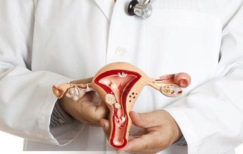
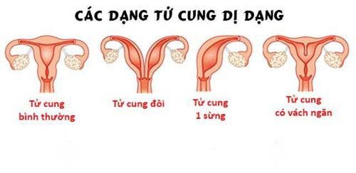
Uterine malformation is one of the congenital malformations that can be encountered in women, uterine malformation affects the ability to conceive and give birth in women.Some types of uterine malformations can be encountered such as:
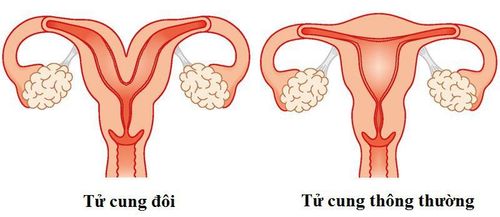
Double uterus or double chambered uterus: Two uteruses can be seen in the pelvis. Separated uterus: This is the most common type of uterine malformation among uterine malformations, accounting for about 40%. Bicornuate uterus: This condition is for the volume of the uterus to be reduced, if it is mild, there is still a possibility of normal pregnancy but increases the rate of premature birth. Unicornuate uterus: In this case, the patient usually has only one ovary and one fallopian tube. Lower chance of pregnancy. Uterine malformation surgery is surgery to correct the uterus, cut the septum, remove the dysfunctional uterus... Helping women to become pregnant and give birth more easily.
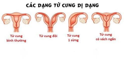
Một số dạng dị dạng tử cung thường gặp
2. Endotracheal anesthesia technique in uterine malformation surgery
Endotracheal anesthesia for uterine malformation surgery is a technique of general anesthesia with endotracheal intubation for the purpose of breathing control during surgery and postoperative resuscitation.2.1 Designation
Non-functional hysterectomy in double uterus, septum removal, repair of malformed uterus... Controlling the airways with a mask is difficult. Maintain anesthesia with inhalational anesthetic.2.2 Contraindications
Patients with medical conditions unable to tolerate surgery. The patient disagreed. Insufficient facilities for anesthesia and resuscitation and inadequate technical skills of the anesthesiologist.3. Endotracheal anesthesia procedure
3.1 Preparation
Performer: Including a team of doctors and nurses in the department of anesthesiology and resuscitation.Equipment for performing the technique required:
Anesthesia machine system with ventilator; balloon oxygen source; monitor vital functions including electrocardiogram, arterial blood pressure, SpO2, EtCO2, temperature, breathing rate; defibrillator, phlegm aspirator. Laryngoscope, endotracheal tube of different sizes, suction tube, mask, bulb, oropharyngeal cannula, Magill pliers, soft mandrin. Medicines: Prepare drugs used in anesthesia and resuscitation and handle complications if any. The necessary means of precaution in case of difficult intubation include: Cook tube, laryngeal mask, flexible bronchoscope, mouth opening pliers, tracheostomy kit...

Thuốc an thần được chỉ định với trường hợp bệnh nhân bị kích động
Full pre-anesthesia examination, detection and prevention of possible complications. Advice and explanation of possible complications in anesthesia and surgery to remove ovarian tumors or remove ovarian tumors. Risk assessment of cases at risk of difficult intubation. Use sedation the night before surgery if needed.
3.2 Steps to take
Patient:Patient position: Lie on back, breathe 100% oxygen 3-6 l/min at least 5 minutes before induction of anesthesia. Install a vital sign monitor, if the patient has laparoscopic ovarian surgery, the EtCO2 parameter must be provided. Establish an effective infusion and administer pharmacological pre-anesthesia. Anesthesia:
Anesthesia can be initiated by intravenous anesthetic, inhalation or a combination of the two. Combine some drugs to increase the effect of anesthetics such as: opioid pain relievers (morphine, fentanyl..), muscle relaxants if necessary. Oral intubation: The condition for intubation is that when the patient is in deep sleep, the degree of muscle relaxation is sufficient in most cases.
Open the mouth, put one hand under the neck to help straighten the neck, then put the laryngoscope to the right of the mouth, move the patient's tongue to the left, push the light deeply, coordinate with the right hand to press down on the cricoid cartilage and find the lid glottis, orifice. Perform rapid induction of anesthesia and the Sellick maneuver in cases of full stomach. Pass the endotracheal tube gently through the glottis, stopping when the balloon of the endotracheal tube passes about 2-3 cm across the vocal cords. Remove the laryngoscope and inflate the endotracheal balloon.
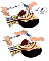
Kỹ thuật đặt nội khí quản đường miệng trong gây mê nội khí quản
Monitoring:
Need to control breathing with a ventilator or hand-held oxygen source. Monitoring the depth of anesthesia based on heart rate, arterial blood pressure, sweating, tearing... Monitor signs and prevent cases of endotracheal intubation in the wrong position, folding...
3.3 Standards for extubation
When the surgery is completed, the endotracheal tube should be extubated when the following criteria are met:The patient is awake, following orders; Lift head for more than 5 seconds, TOF > 0.9 (if any); Spontaneous breathing, breathing rate within normal limits; Stable pulse and blood pressure; Body temperature > 35 degrees Celsius; There were no complications from anesthesia and after surgery.
4. Complications and management of complications due to intubation
4.1 Reflux of gastric juice into the airway
Signs: There is digestive juices in the oral cavity and airways. Treatment: Drain the fluid, place the patient in a low lying position, tilt the head to one side; Rapid intubation of the endotracheal tube and aspiration of the airway fluid; Monitoring and prevention of lung infections after surgery
Người bệnh cần được theo dõi để tránh nhiễm trùng phổi sau phẫu thuật
4.2 Hemodynamic disorders
Hypotension or hypertension, arrhythmia (may be bradycardia, tachycardia, arrhythmia). Treatment depends on the signs and causes.4.3 Complications due to intubation
Intubation failure: Re-intubate following difficult intubation procedures or switch to another method of anesthesia. Misplaced in the stomach: Signs: Auscultation of the lungs without alveolar murmurs, no measurement of EtCO2. Treatment: Reinsert the endotracheal tube. Vocal, air, and bronchial spasms Difficult or impossible to ventilate the lungs due to spasm, auscultation with rales or muted lungs. Management: Provide adequate oxygen, add sleeping pills and muscle relaxants, ensure ventilation, give bronchodilators and corticosteroids; If breathing is not controlled, a difficult intubation procedure should be used. Injuries when inserting a tube include: Bleeding, broken teeth, damage to the vocal cords, falling foreign bodies into the airways...Need to be handled depending on the injury.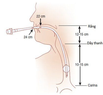
Trường hợp bệnh nhân không đặt được nội khí quản cần chuyển sang phương pháp vô cảm khác.
4.4 Respiratory complications
The endotracheal fold and retraction cause the endotracheal tube to be pushed deep into one lung, collapse or open the respiratory system, running out of oxygen...Treatment: Need to immediately ensure ventilation and provide 100% oxygen, resolve the cause.
4.5 Complications after extubation
Respiratory failure . Sore throat hoarse voice. Constriction of the larynx, trachea and bronchi. Inflammation of the upper respiratory tract. Narrow larynx, trachea. Treat the patient on a case-by-case basis and on a case-by-case basis. Treat the patient on a case-by-case basis and on a case-by-case basis.Uterine malformations reduce or cause infertility in women. Uterine correction surgery increases the chances of pregnancy. Anesthesia and surgery can both cause some complications, so it is necessary to choose reliable addresses, with a team of highly specialized doctors, and modern machinery to help limit the risk of accidents.
Vinmec International General Hospital is one of the hospitals that not only ensures professional quality with a team of leading medical doctors, modern equipment and technology, but also stands out for its examination and consultation services. comprehensive and professional medical consultation and treatment; civilized, polite, safe and sterile medical examination and treatment space. Customers when choosing to perform tests here can be completely assured of the accuracy of test results.
Master. Dr. Ton That Quang has more than 15 years of experience working in the Anesthesia - Resuscitation industry. Doctor Quang was a doctor at the Department of Anesthesiology and Resuscitation at Khanh Hoa General Hospital and a lecturer at the provincial level of Emergency Resuscitation in Obstetrics and Gynecology before being an Anesthesiologist and Resuscitation Doctor at the Department of General Surgery, General Hospital. Vinmec Nha Trang International.
Customers can directly go to Vinmec Health system nationwide to visit or contact the hotline here for support.


