This is an automatically translated article.
Articles are professionally approved by Master. Doctor Nguyen Quang Duc - Doctor of Nuclear Medicine - Department of Diagnostic Imaging and Nuclear Medicine, Vinmec Times City International Hospital.Bone scintigraphy uses nuclei that emit radiation, detected by a scanner, to create images of bones. This method helps find bone cancer or identify cancer from elsewhere that has spread to the bone. Before the bone scan, do you have to fast or not is a question many people are interested in.
1. What is a bone scan?
Bone scan is a nuclear medicine imaging test that helps diagnose and monitor bone diseases. Your doctor may order a bone scan for someone with unexplained bone pain, infection, or bone injury that can't be seen on an X-ray. It is also an important method for detecting cancer that has spread to the bones , such as breast or prostate cancer . Although not specific, but bone scintigraphy has high sensitivity, is the only technique to evaluate the physiological, metabolic and metabolic state of the musculoskeletal system. Thanks to its ability to scan the entire skeleton, this method becomes useful when diagnosing many bone disorders, including:
Fractures Arthritis Arthritis Paget's disease of bone (abnormal bone structure disorder) Primary bone cancer Cancer metastases to bone Monitor for joint infection and osteomyelitis after joint or bone replacement Avascular necrosis, metabolic disease causing osteoporosis, osteomalacia, etc. Evaluation of response to chemotherapy, radiotherapy, antiretroviral therapy biopsies or other methods Determine the site for puncture, bone biopsy. Although bone scintigraphy produces images on a radiograph, there is very little radiation exposure, even less than with a CT scan (computed tomography). This method does not cause any side effects or complications. Most radioactive tracer will clear from the patient's body within 24 hours, a few can persist for 3 days.
MORE: Is bone scan dangerous?
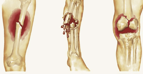
Phương pháp xạ hình xương giúp chẩn đoán nhiều vấn đề về xương
2. Bone scintigraphy procedure
2.1. Before getting into work
Before the bone scan, do you have to fast or not is a question that many patients care about. According to doctors, you do not need to limit your food intake or avoid certain activities before having a bone scan. If you are taking medication, especially one containing bismuth (Pepto-Bismol), or if you have had a barium X-ray within the past 4 days, tell your doctor. Because barium and bismuth have the potential to affect bone scan results.
Immediately prior to the procedure, the patient is often asked to remove jewelry or other metal objects. Some cases require you to empty your bladder before starting the scan. A full bladder can block the view of the pelvis.
Bone scintigraphy will be limited to pregnant or lactating women because of concerns about the risk of radiation exposure to the fetus. Therefore, it is necessary to inform your doctor if you are or may become pregnant, as well as are breast-feeding.
2.2. While doing
As mentioned, this is a procedure in the Department of Radiology and Nuclear Medicine. The patient will be injected with a small amount of radioactive tracer into a vein depending on the location to be scanned.
The areas of the body where cells and tissues are in the process of "self-repairing" will take up the most tracer. From there, radiographs will highlight these areas, showing abnormalities related to disease or trauma.
In general, the bone scan procedure consists of 2 main stages:
Injecting the tracer The radioactive tracer is injected into a vein in the patient's arm. The time and dose of injection will vary depending on the indication for performing the bone scan. In some cases, it is possible to record images immediately after injection. However, the main images are taken 2 - 4 hours later, when the tracer has circulated and absorbed into the bone. Patients are often advised to drink plenty of fluids in the meantime.
Scanning The doctor will ask the patient to lie still on the scanning table connected to a specialized camera device that scans the body. This phase can last up to 1 hour, but is not painful for the patient. Three-phase bone scintigraphy is a technique that records a series of moving images at different times, used to evaluate osteomyelitis or connective tissue inflammation. 3 specific phases include: perfusion phase, blood lake phase and late phase 3 hours after injection.
To see more clearly some types of bones in the body, the doctor may continue to assign the patient a single photon emission computed tomography (SPECT) scan. This method is capable of imaging diseases that are particularly localized deep in the bone or where it is difficult to see through bone scintigraphy alone.
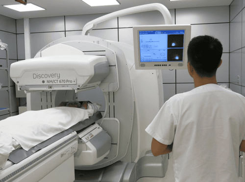
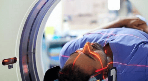
Quy trình thực hiện của phương pháp xạ hình xương
2.3. After doing
In general, bone scans are not associated with side effects, nor do they require any follow-up care. The radiation from the scanners usually wears off completely within 2 days of completing the scan.
MORE: Is it necessary to isolate after bone scan?
3. Bone scan results
The radiologist or technician will read the patient's scan results to look for abnormalities in the bones. Results are considered normal when the radiotracer image is spread throughout the body. Abnormal results are when visible dark spots appear on the scanned image. These can be disorders in the bones, such as cancer or arthritis, a bone infection.
Characteristic of metastatic cancer to bone is the appearance of many foci, poles account for about 80%, remaining about 10% metastasize to skull and 10% to long bones. When there is metastasis, on the bone scan, the whole bone will be abnormally dark and irregularly irradiated. Where there is bone destruction, there will be foci of reduced radioactivity. When an abnormal focus is detected on the scan, the patient may be asked to take X-rays of those bones to confirm the diagnosis of metastases.
Based on the bone scan results, the doctor also decides whether to have amputation surgery for bone sarcoma. Bone scintigraphy gradually becomes a screening method for disease at the time of initial diagnosis. If you still have questions about bone scans, please talk to a specialist for the best advice and support.
In order to perform bone scintigraphy for clear and accurate diagnosis results, currently, Vinmec International General Hospital has introduced the most modern SPECT/CT Discovery NM/CT 670 Pro system with 16-sequence CT. of the world's leading medical equipment company GE Healthcare (USA), for high quality images, helping to diagnose early bone diseases to be investigated. Accordingly, the entire examination and imaging process at Vinmec is performed by a team of experienced medical staff, well-trained at home and abroad, so they can advise and support maximum. for customers during the shooting process, even for foreign customers.
Please dial HOTLINE for more information or register for an appointment HERE. Download MyVinmec app to make appointments faster and to manage your bookings easily.