This is an automatically translated article.
The article is professionally consulted by Dr., Doctor Tran Nhu Tu and Master, Doctor Le Thi Hong Vu - Department of Diagnostic Imaging - Vinmec Danang International Hospital.
Chest X-ray is a common appointment of doctors when they want to check their health and screen for lung diseases. The article will provide you with information on how to perform, purposes, and requirements to help you better understand this test.
1. What is a chest X-ray?
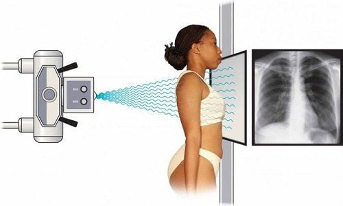
Chest X-ray is a technique that uses an X-ray machine in a special room with a movable X-ray tube attached to a large metal rod, the patient will be instructed to stand in front of a plate containing X-ray film. or a special receiver that can record images of the heart, lungs, airways, blood vessels, and lymph nodes.In order to fully explore the lungs, taking a chest X-ray is only a necessary first step, but it does not bring the most authentic results. People can clearly see based on some of the following pros and cons.
Advantage: Fast. Less money. See the whole lungs, heart shadow, ribcage. Large, unobstructed lesions are seen on both lungs. Cons: No small or early-stage lesions can be seen on both lungs. Lung lesions can be covered by the shadow of the heart, ribs, etc. It is difficult to detect lesions at hard-to-see locations such as the two apex of the lung. The internal characteristics of the lesion are not clearly visible.

Hình ảnh chụp X - quang phổi
2. What disease can be detected by chest X-ray?
When taking an X-ray, the X-ray machine will project chest X-rays to produce specific images. X-rays penetrate soft tissues, body fluids, and film to produce images of the lungs, heart, blood vessels, and structures of the chest wall such as ribs...Based on this image, the doctor can check Investigate, identify and diagnose some of the following:
Determine if there is fluid or air inside the pleural cavity or peripulmonary space; Diagnosing whether the patient has heart and lung diseases such as: Pneumonia, atelectasis, cancer, etc. If traumatized, it can be determined that the ribs are broken or damaged.
3. When should a chest X-ray be taken?
Although there are many limitations, but conventional chest X-ray is still the first method to help doctors diagnose the disease. Compared with other health examination and pathological diagnostic techniques, chest X-ray is applied in many different cases:Checking lung status during periodic health check-ups. Indicated when the patient has symptoms such as: Shortness of breath, chest pain, trauma, persistent cough, .. Diagnosis and screening pathology if suspected at risk of chest trauma, contusion, pneumonia, pulmonary tuberculosis. , lung tumor, pleural effusion,... Detecting lung abnormalities as well as monitoring progress in case of lung disease. Severe pain after an injury or heart disease.

Chỉ định chụp X-quang phổi khi khám sức khỏe định kỳ
4. Routine chest X-ray procedure
The X-ray room is specially designed with equipment installed to prevent X-rays from escaping and affecting other patients. For most hospitals, medical facilities share a common process that includes the following steps.Move to the imaging room as directed by either the doctor or the technician. You will be instructed to stand near a sheet of X-ray film inside or an instrument with a special receiver that records the images into a computer. Next, you are instructed how to stand and hold your breath to increase the sharpness of the X-ray image. Once the capture is done you will be able to return to normal activities. Your doctor will give you an explanation of your condition as soon as the X-ray results are available. Also, let you know if you need to take any additional steps.
During the imaging process, X-rays can affect the patient's body. But you are completely assured when exposed because the amount of X-rays is very low, even lower than the X-rays that you get infected when going out on the street. Therefore, there will be no significant harm.

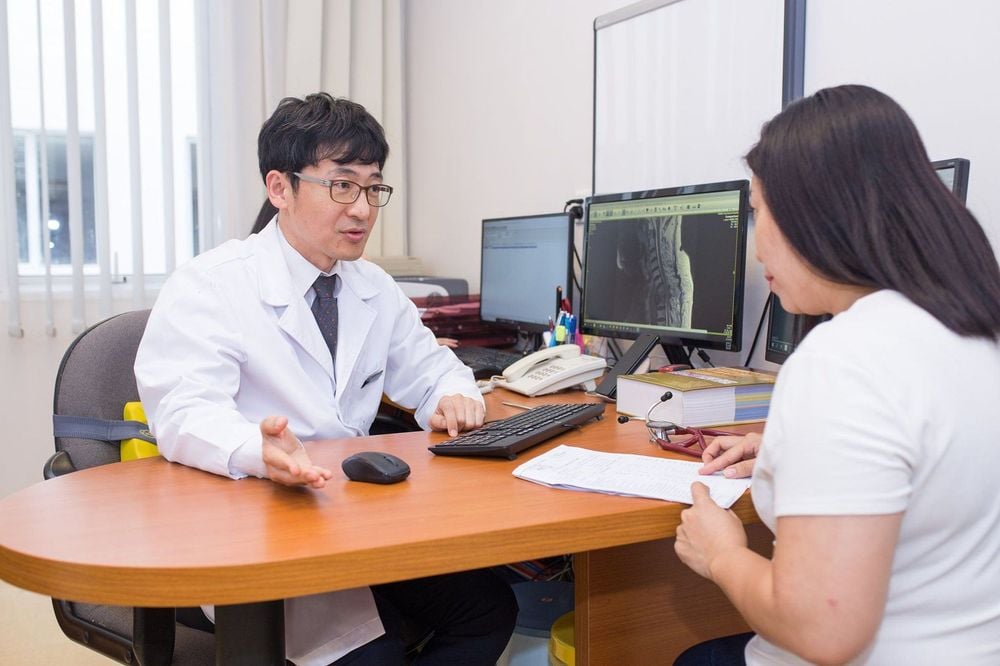
Kết quả chụp X-quang sẽ được bác sĩ chuyên khoa phân tích và đưa ra kết luận
5. Instructions for reading chest X-ray results
Patients will be able to see their condition through chest X-ray films. Specifically, a normal result will have:Normal lung size and shape. There were no masses or complications in the lung, the pleura was normal. The blood vessels and heart tissue are also of normal size. The rib cage and spine are also normal. No accumulation of liquid or gas, foreign body in the lung cavity was detected. Abnormal results show that you are suffering from related diseases:
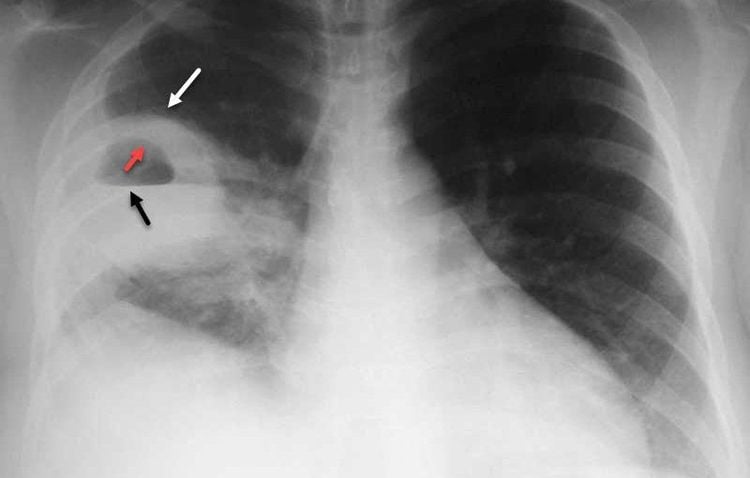
Detect tumors, trauma, pulmonary edema. See the inflammation of pulmonary tuberculosis, pneumonia. Detect fluid around the lungs, surrounding air. See damage to ribs, collarbone. Detects enlarged lymph nodes or foreign bodies in the airways, esophagus, or lungs. If you have any questions, you can consult with your doctor to better assess your lung condition.

Hình ảnh kết quả x-quang phổi bất thường
6. What should be noted when taking X-ray of the lungs?
Chest X-ray does not cause pain, but to ensure accurate results, patients should note a few things:Should bring summary of medical records, test sheets, X-ray results before that if any so that in some cases doctors need a comparison to draw conclusions. If you are a female patient, you should inform your doctor if you suspect that you are pregnant or are pregnant to avoid the adverse effects of X-rays on the fetus. Patients need to wear thin, light clothes or gowns of medical examination facilities. Remove objects that affect the photographic film such as rings, rings, hairpins, glasses, ...
7. How much does a chest X-ray cost?
The cost of chest X-ray is also a question that people are interested in. According to the decision of the Ministry of Health, the minimum price for taking pictures will be about 95,000 VND / time. Abroad, the cost of a lung scan is from 54 to 204 USD, with an average of 111 USD. This price does not include other test and treatment indications. Health insurance use cases will also not be covered for these chest X-ray tests.Depending on different medical facilities and hospitals, the price of X-ray has different fluctuations, possibly higher than the regulations of the Ministry of Health. This is a completely low fee compared to other modern diagnostic methods. Therefore, you should refer to the X-ray first and then, if indicated, further tests may be performed.

Định kỳ kiểm tra sức khỏe giúp bạn phát hiện sớm bệnh lý
Above is information about chest X-ray technique and hope to answer your questions about the question "Diseases that can be diagnosed early by chest X-ray". Always pay attention to regular health check-ups to detect dangerous cardiopulmonary diseases early. From there, a timely treatment solution can be obtained.
Dr. Tran Nhu Tu with over 20 years of experience, of which 3 years as Deputy Head of the Paraclinical Department - Hoa Vang Hospital, 16 years as Deputy Head of Imaging Department at Da Nang Hospital, 4 years as Head of Department Diagnostic imaging at Da Nang Obstetrics - Pediatrics Hospital; Dr. Tu makes accurate diagnoses, minimizes errors based on acquired images, and is ready to serve patients 24/24 in emergencies and emergencies.
Dr. Tu has participated in domestic and foreign training courses such as: Master of Radiology, Research Fellow in Diagnostic Imaging at Hanoi Medical University, Diagnostic Imaging at CHU Reims - France, Electricity interventional radiology at Winterthur hospital - Switzerland, interventional radiology at Singapore General Hospital... before being the head of the Department of Diagnostic Imaging at Vinmec Danang Hospital as it is now.
Master doctor Le Thi Hong Vu has good English, has participated in many short-term and continuous training courses on annual diagnostic imaging. The doctor has more than 7 years of experience as a doctor in the field of diagnostic imaging, especially ultrasound.
To register for chest X-ray at Vinmec International General Hospital, you can contact Vinmec Health System nationwide, or register online HERE.
MORE:
Find out information about empyema Causes and diagnosis of lung abscess How is lung cancer diagnosed?


