This is an automatically translated article.
Atrioventricular canals are congenital abnormalities of the heart that are associated with Down syndrome. Diagnosing atrioventricular canal congenital heart disease helps children detect the disease early and treat it.1. What is the congenital atrioventricular canal?
The atrioventricular canal is a type of congenital anomaly that causes damage to the structure from the endothelium of the heart during the fetal period.The endothelium is a germ of the endothelium, appearing in the 4th week of pregnancy, responsible for creating the primary atrial septum, the ventricular septum and the permanent atrioventricular valves for the heart.
Atrioventricular canal has 2 types of disease: partial atrioventricular channel and partial atrioventricular channel.
2. Diagnostic measures for atrioventricular canal congenital heart disease
2.1 Diagnosis of total atrioventricular canal The work, measures to diagnose total atrioventricular canal include:Symptoms of total atrioventricular canal usually occur early, when the child is less than 1 year old, most of the cases below 6 months old had obvious symptoms such as:
Tiredness, shortness of breath. Weak suckling, malnutrition, growth retardation. Recurrent lung infections. Rapid breathing, pulling, slight purple lips on exertion. 2.2 Subclinical diagnosis
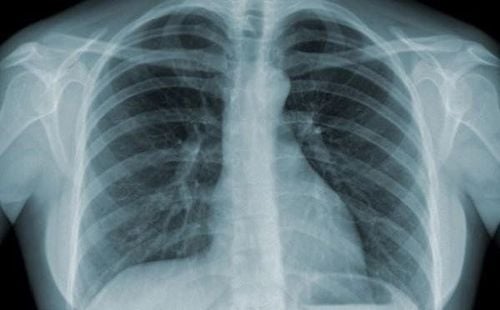
Chẩn đoán XQ tim phổi
Cardiopulmonary X-ray: The whole heart is enlarged, especially the right atrium, increasing active pulmonary circulation. ECG: Left ventricular axis deviation or in an irregular region, right ventricular dominant biventricular enlargement. Large right or left atrium or both atria. Long PR such as 1st degree AV block, incomplete right bundle branch block. Echocardiography: Receptive ventricular septal defect, primary atrial septal defect, mitral valve injury. Assess flow, pulmonary artery pressure, ventricular function, and chamber size. 2.3 Diagnostic methods of partial AV channel The diagnostic methods of partial AV channel are symptomatic diagnosis and subclinical diagnosis (pulmonary X-ray, EEG and echocardiography). Diagnostic symptoms in each type of partial atrioventricular channel are different.
Primary atrial septal defect Symptoms are usually very few, except in the case of significant mitral regurgitation accompanied by early heart failure in children.
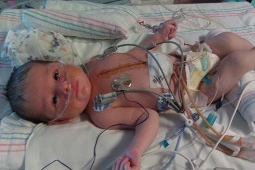
Trẻ bị kênh nhĩ thất có thể kèm suy tim sớm
ECG: Similar to the total atrioventricular channel, left ventricular axis deviation or in an indeterminate region, right ventricular dominant biventricular enlargement. Large right or left atrium or both atria. Long PR such as 1st degree AV block, incomplete right bundle branch block.
Monoatrial heart The clinical and laboratory findings of this type are similar to those of the primary septal defect. However, there is often an additional mild cyanosis, more often with exertion.
In addition, there is often associated systemic venous abnormalities and structural abnormalities of the heart.
Ventricular septal defect Similar to ventricular septal defect, the membranous part is located high above the interventricular septum associated with mitral or tricuspid valve damage.
Clinical signs are similar to ventricular septal defect.
Especially, when the ECG is diagnosed, the left axis of the heart is deviated with increased right ventricular load.
Partial atrioventricular canal with left ventricular–right atrioventricular septal defect The flow passes from the left ventricle to the right atrium with damage to the spleen of the tricuspid valve.
Clinical: Similar to ventricular septal defect with pulmonary hypertension occurring early in infancy.
Diagnosis of ECG shows: left axis, long PR, right bundle branch block, atrial enlargement, left ventricular or biventricular hypertrophy.
The definitive diagnosis method is based on echocardiography.
3. Complications of congenital heart disease atrioventricular canal
The principle of treatment of atrioventricular canal congenital heart disease is surgical repair of defects and prevention of dangerous complications. Therefore, the earlier the treatment time, the more the child can prevent dangerous complications that are difficult to overcome when they grow up.
Cần sớm chẩn đoán phát hiện kênh nhĩ thất
Pneumonia
If the atrioventricular canal is not treated, it can lead to pneumonia - a recurrent serious lung infection.
Dilates the chambers of the heart
Increased blood flow through the heart forces the heart to work harder than usual, causing dilation.
Heart failure
If left untreated, the atrioventricular canal often leads to heart failure - a condition when the heart cannot pump enough blood to meet the body's needs.
Pulmonary hypertension
When the left ventricle weakens and can no longer pump enough blood, the pressure increases through the pulmonary veins to the arteries in the lungs, causing pulmonary hypertension.
Measures to diagnose atrioventricular canal congenital heart disease help parents and families to detect the child's disease early, have treatment and prevent complications early.
Please dial HOTLINE for more information or register for an appointment HERE. Download MyVinmec app to make appointments faster and to manage your bookings easily.


