This is an automatically translated article.
The article is professionally consulted by Specialist Doctor II Nguyen Khanh Nam - Doctor of Odonto-Stomatology - Department of General Surgery - Vinmec Nha Trang International General Hospital.
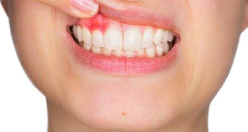
Periodontitis is a common oral disease that causes pain and affects patients' daily activities. The diagnosis and treatment of periodontitis should be implemented early and properly to avoid leading to other complicated complications.
1. Diagnosis of periodontitis
1.1 For acute periodontitis definite diagnosis Clinical signs help doctors diagnose acute periodontitis:Whole body: Patient feels tired, has a high fever ≥ 38 ̊C, accompanied by signs of infection Infections such as dry lips, dirty tongue, may have a ganglion reaction in the area under the jaw or under the chin. Function: Occurrence of toothache with natural, continuous, intense pain, pain spreading to migraine, increasing degree of chewing, little response to analgesics, patients can define themselves clearly painful tooth location. In addition, patients with acute periodontitis also have a feeling of tooth protrusion, tooth pain while biting and eating food, making the patient not dare to chew. Physical symptoms: Corresponding to the location of the damaged tooth, the outer skin is swollen, red, without clear boundaries, painful when pressed, and there are corresponding lymph nodes. Teeth can be discolored or not discolored, when examined often damage caused by caries that have not been sealed, or teeth that have been treated, or other injuries that are not due to caries. In addition, there may be obvious signs of loose teeth, usually grade 2 or 3, and when percussion along the teeth, the pain is more intense than in horizontal percussion. The gingival mucosa is swollen, red, painful, and the tissue is loose. If a pulp test is performed, the electrical and thermal test results are negative because the pulp is necrotic. A number of subclinical signs help doctors diagnose acute peri-pedunculitis:
X-ray: A blurred image of the pedicle can be obtained, the border is not clear, and there are signs of stretching of the ligament around the pedicle. Blood tests: The number of neutrophils increases, the erythrocyte sedimentation rate increases... Differential diagnosis Periodontitis should be differentiated from acute pulpitis:
| Triệu chứng | Viêm tủy cấp | Viêm quanh cuống cấp |
| Dấu hiệu toàn thân | Không có dấu hiệu toàn thân | Bệnh nhân mệt mỏi, sốt cao, có phản ứng hạch vùng. |
| Đặc điểm cơn đau | Đau tự nhiên, thành cơn, cơn đau hay xuất hiện về đêm, đau tăng khi nhai thức ăn. | Đau tự nhiên, âm ỉ, liên tục,răng có hiểu hiện lung lay, chồi cao. |
| Gõ dọc | Đau ít | Đau nhiều |
| Thử tủy | (+) Dương tính | (-) Âm tính |
| Chụp X-quang | Vùng cuống cho hình ảnh bình thường |
Có hình ảnh dãn rộng dây chằng |
Whole body: Patient has malaise, headache, mild fever below 38 ̊C or sometimes not fever. Functional symptoms: Dull, continuous pain in the damaged tooth, feeling that the tooth is high, the pain increases when the two jaws touch. Physical symptoms include: The corresponding gingival gap in the affected tooth area is mildly swollen, red, full, and painful when pressed. However, less swelling is seen in the corresponding external skin, possibly with small mobile lymph nodes. Teeth are gray or not. In addition, subacute periodontitis causes carious lesions on the tooth surfaces, the teeth are loose at grade 1, 2, if you tap vertically, you will feel more pain than when you tap horizontally. Marrow test was negative. In terms of subclinical, on the X-ray, you can see a blurred image of the pedicle, and a slight stretch of the ligament in the pedicle.
Differential diagnosis Signs to help differentiate acute periarthritis:
| Triệu chứng | Viêm quanh cuống cấp | Viêm quanh cuống bán cấp |
| Dấu hiệu toàn thân | Bệnh nhân mệt mỏi, sốt cao, có phản ứng hạch vùng. | Bệnh nhân khó chịu, đau đầu, sốt nhẹ dưới 38˚C hoặc không có sốt. |
| Đặc điểm cơn đau | Đau tự nhiên, âm ỉ, liên tục, biểu hiện răng lung lay, chồi cao. | Đau âm ỉ, liên tục ở vị trí răng tổn thương, cảm giác răng chồi cao, đau tăng khi hai hàm chạm nhau. |
| Gõ dọc | Đau nhiều | Đau ít |
Function: Only history of pain from episodes of acute myelitis, acute periradicular inflammation, or acute periradicular abscess. Physical symptoms: Notice the change in tooth color, opaque gray color in the dentin, partly reflected through the enamel layer. The corresponding gingival recess around the apex may be slightly swollen, with fistulas or fistula scars at the apex. Sometimes the fistula does not appear in the oral cavity but outside the skin or the base of the nose depending on the location of the cyst and the abscess. When percussion, the patient does not feel pain or only mild pain in the root area. In addition, teeth may become loose when alveolar bone resorption occurs. Marrow test was negative. When taking X-rays for laboratory review, he noticed:
When putting gutta-percha through the fistula in the mouth, the doctor can see the image of the source of the pus on the X-ray film. Chronic periradicular abscess: The image of bone resorption is not defined. Granulomas and cysts: When bone resorption is clearly demarcated. For chronic periarthritis, it is not possible to make an accurate differential diagnosis without a biopsy.
Once the correct diagnosis has been made, the doctor can decide to choose the right treatment for the root canal inflammation for each specific case.
Đối với viêm quanh cuống mạn, không thể chẩn đoán phân biệt chính xác các thể bệnh nếu không thực hiện sinh thiết
2. How to treat inflammation of the tooth root
2.1 Principles of treatment of root canal inflammation Remove all infected and necrotic tissue inside the root canal. Good drainage for inflammatory tissue in the pedicle. Seal the root canal system, creating optimal conditions for the recovery of the stem tissue. If the prognosis of endodontic treatment is not effective, surgery should be indicated for the patient's root canal. 2.2 Treatment protocol for periodontal inflammation Teeth with acute or subacute periodontitis: drainage of pulp chamber. Then use antibiotics effective against anaerobic and Gram (-), combined with pain relief, improve physical condition to conduct endodontic treatment.3. How to cure specific root inflammation
3.1. Systemic treatment For painful diseases (acute periapical inflammation, acute periapical abscess), treatment of aphthous stomatitis with systemic antibiotics is required, especially in the case of patients with acute periapical abscess. accompanied by cellulitis.3.2. Endodontic treatment Clean and shape the root canal system. Place Ca(OH)2 in the root canal, in order to neutralize inflammatory tissue in the pedicle, and disinfect the root canal system. Seal the root canal system. Perform crown restoration. 3.3. Surgical treatment Treatment of apical inflammation by surgery is applied in the following cases:
After endodontic treatment, there are irreversible peri-apical lesions due to various causes. After the cause tooth and related teeth have been endodontically treated, but the apical lesion has not progressed well, surgical treatment must be carried out to remove the entire capsule shell, which may or may not be present. do not cut the root of the root cause. If there is a root canal cut, the doctor must conduct a reverse root canal.
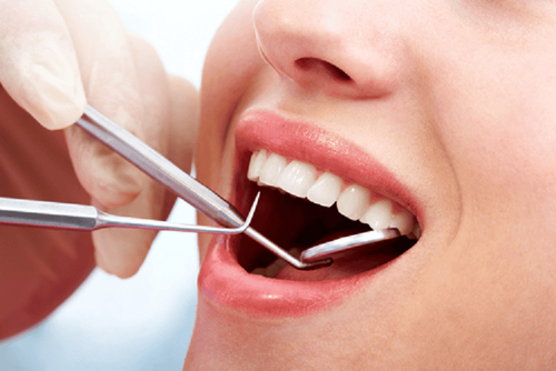
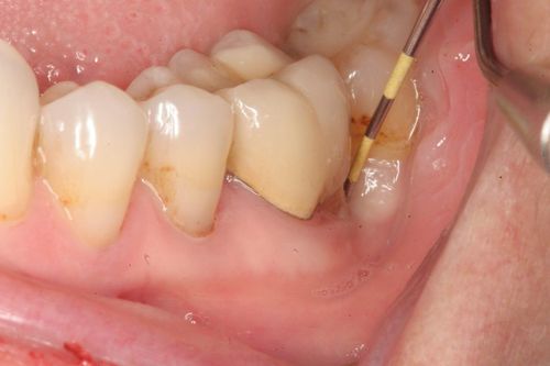
Điều trị nội nha chữa viêm cuống răng
4. Treatment of periodontitis at Vinmec Hai Phong International Hospital
Currently, at Vinmec Hai Phong International General Hospital, the technique of root canal treatment and sealing of the root canal system with cold Gutta percha has been applied using a rotating file.Although root canal treatment is a technique that has been around for a long time, but treatment with file machines has only been born in the past few years. Currently, this new technology has been approached and applied by Vinmec to oral health care, along with a system of modern machinery and equipment, ensuring the best quality of medical examination and treatment for our patients:
System KAVO dental chair system (Germany). Camera at the tooth chair. Panoramic camera Gendex brand. In situ alveolar X-ray machine, Gendex Phosphorus film scanner. Dently ultrasound machine. Radii Plus teeth whitening lamp, made in Australia, endodontic machine, Root canal length meter, Root canal treatment uses a measuring device to determine the length of the foot to help the dentist work to the full working length. The machine file protaper helps to clean and shape the canal well, saving work time, and the customer does not have to open his mouth for a long time compared to when doing manual files. The apex locator X-ray machine helps to examine the artificial pulp before, during and after welding.
BSCK II Nguyen Khanh Nam has more than 30 years of experience in Odonto-Stomatology. Doctor Nam was formerly Head of Odonto-Stomatology Department of Khanh Hoa Provincial Hospital and Deputy Director of Service Center of Khanh Hoa Provincial Hospital before being a General Internal Medicine Doctor of Medical Examination and Internal Medicine Department of Vinmec Nha Trang International General Hospital. like nowadays.
Please dial HOTLINE for more information or register for an appointment HERE. Download MyVinmec app to make appointments faster and to manage your bookings easily.