This is an automatically translated article.
The article is professionally consulted by Master, Doctor Pham Van Hung - Department of Medical Examination & Internal Medicine - Vinmec Danang International General Hospital.
Transposition of the great arteries is a rare but serious congenital heart disease. Symptoms appear at birth, in which the two main arteries that carry blood out of the heart are reversed.
Normally, the pulmonary artery and the right ventricle communicate with each other, the aorta and the left ventricle are connected. But with transposition of the great arteries, it is different: the pulmonary artery takes the place of the aorta in the left ventricle; Similarly, the aorta and right ventricle are joined together. It is a severe disease, with a mortality rate of 30% in the first week of life, 50% in the first month, and 90% in the first year.
1. What is transposition of the great arteries?
Transposition of the great arteries is in the group of diseases "abnormal connection of the ventricle and the main artery", including transposition of the great arteries, the common trunk of the great arteries, and the right ventricle with two outflow tracts.The disease requires early diagnosis, the mortality rate is high with about 90% of children dying before 1 year of age if not operated. The 5-year survival rate in children with early detection and surgery is over 80%. Therefore, it is best to be detected and diagnosed during pregnancy.
2. Symptoms of transposition of the great arteries

Symptoms of severe heart failure: Shortness of breath, edema, palpitations, irregular heartbeat. Patients without symptoms of heart failure often present with severe cyanosis. There may be brain abscess, cerebrovascular accident. Physical symptoms:
Normal blood pressure, normal pulse. Chest deformity. T1 is normal. T2 alone. No murmur or systolic murmur (a murmur heard due to blood flow through the narrowing).
3. Subclinical diagnosis of transposition of the great arteries
3.1 Electrocardiogram Record the electrical activity of the heart, from which abnormal changes in the structure of the heart can be seen. Diagnosis is usually nonspecific with:Usually sharp, high P waves (presenting right atrial thickening). When the amount of blood to the lungs is high, but the atrial septal defect is small (botal hole), there is left atrial thickening. Common right ventricular thickening, sometimes with 2 ventricular thickening. The T waves are often deep inversion in the right thoracic leads. 3.2 Chest X-ray is ovoid if there is a lot of blood to the lungs. Depending on the stenosis of the pulmonary artery, the pulmonary circulation will increase or decrease. The great artery bundle is often narrow due to the anterior and posterior position of the aorta and pulmonary artery. The radiograph showed all 3 images: ovoid heart, increased pulmonary circulation, and narrow great artery bundle suggesting transposition of the great arteries.
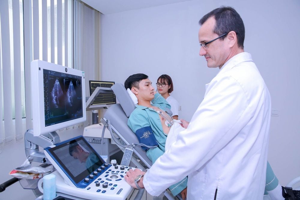
3.3 Echocardiography Uses the transformation of ultrasound waves recorded on a sensor to create dynamic images of the heart and its valves, thereby assessing the structure and function of the heart.
This is the standard to help doctors orient and treat transposition of the great arteries.

4. Treatment of transposition of the great arteries
4.1 Medical treatment Intravenous Prostaglandin: To keep the ductus arteriosus open (one of the vital shunts that help blood flow from the pulmonary artery to the aorta). From there, it helps to connect the two circulations, improving the oxygen levels in the blood to the organs until the patient can perform surgery.Atrial septal defect: Similar to the Prostaglandin transmission mechanism, atrial septal defect will create a connection between the two circulations through the two atria. Thanks to this communication, oxygen-rich blood from the left atrium enters the right atrium, following the aorta to the organs that nourish the body.
4.2 Surgical treatment There are two surgical directions, which are arterial bypass surgery and atrial displacement surgery:
Arterial bypass surgery: This is a common surgery to correct the position of the great arteries that bring the arteries to the heart. The vessel returns to its correct position: the pulmonary artery and the right ventricle communicate and the aorta communicates with the left ventricle. This surgery is usually done in the first week after the baby's birth.
Common complications of this method include: coronary artery failure, myocardial ischemia, ventricular dysfunction, arrhythmia,...
Atrial conversion surgery: The principle is to create a tunnel between the two atria. As a result, hypoxic blood flows from the right atrium to the left atrium to the left ventricle and pulmonary artery, and oxygen-rich blood reaches the right ventricle and aorta. Applying this method, will increase the burden of the right ventricle, the right ventricle will perform the function of the left ventricle and vice versa, the left ventricle will bring blood to the lungs to perform gas exchange. Follow this direction, there are the following surgical methods:
Mustard surgery: Create a path by the pericardium, blood from the vena cava will return to the left ventricle and pulmonary artery, blood from the pulmonary vein will return to the right ventricle and aorta. Senning surgery: Same principle as Mustard surgery but uses the atrial septum to block the atrial chambers. These two surgical methods often have late complications. The most common complications were superior vena cava obstruction, inferior vena cava obstruction, and fistula of the anastomosis between the pulmonary vein and the tricuspid fossa. Patients often have right heart failure, tricuspid regurgitation due to increased right ventricular load, arrhythmia, conduction disturbances.
5. Caring for patients with transposition of the great arteries after surgery
After surgery, the survival rate for the first 5 years of life is approximately greater than 80%. After surgery, the patient needs to be cared for and monitored for a lifetime of cardiovascular status. After the patient is stabilized and discharged from the hospital, the mother still needs to closely monitor the child's condition, so she should record the child's diagnoses, medications, and abnormal symptoms. Transposition of the great arteries is a congenital heart disease, which needs to be detected early and promptly. The disease has a high mortality rate of 90% without surgery. Transposition of the great arteries can be detected during pregnancy.Vi so if you have doubts about your baby's condition, you should go to a medical center that specializes in cardiology to examine and treat transposition of the great arteries early. .Master, Doctor Pham Van Hung has 30 years of experience in examination and treatment of internal diseases, especially in Cardiology: coronary arteries, heart failure, heart valves, arrhythmias. ..Master, Dr. Hung used to hold the position of Deputy Head of Internal Cardiology Department and Head of Interventional Cardiology Unit at Da Nang General Hospital and is currently working at Department of Medical Examination and Internal Medicine, Internal Cardiology, and Cardiology. Interventional circuit at Vinmec Da Nang International General Hospital.
Please dial HOTLINE for more information or register for an appointment HERE. Download MyVinmec app to make appointments faster and to manage your bookings easily.


