This is an automatically translated article.
The article is professionally consulted by Master, Doctor Vu Thi Tuyet Mai - Cardiologist - Cardiovascular Center - Vinmec Central Park International General Hospital.Acute right ventricular failure is a complex syndrome, accounting for 3 - 9% of hospitalized patients for heart failure and for 5 - 17% of deaths among hospitalized patients.
1. What is acute right ventricular failure?
The right ventricle has a triangular shape, the structure consists of 3 parts: the receiving chamber, the apex and the funnel. The ventricles are composed of a system of muscle fibers that intertwine in three-dimensional space. Acute right ventricular failure is a clinical syndrome characterized by the inability of the right ventricle to provide adequate blood flow to the pulmonary circulation under conditions of normal central venous pressure. The consequences of the disease lead to right ventricular dilatation and tricuspid regurgitation.Causes of acute right ventricular failure include:
● Extensive pulmonary infarction;
● Acute pulmonary hypertension;
● Acute respiratory distress syndrome (ARDS);
Right ventricular myocardial infarction on ECG;
Infections and infectious cardiomyopathies;
Ventilator;
● After heart-lung transplant surgery;
● After placement of left ventricular assist devices;
● Pericardial disease
● Acute exacerbation on the background of a chronically damaged right ventricle (primary pulmonary hypertension, right ventricular dilatation, right ventricular hypertrophy).
2. Diagnostic method for acute right ventricular failure
2.1 Diagnosis by clinical symptomsSymptoms of acute right ventricular failure include:
● Manifestations of venous stasis and fluid retention
● Hypoxia
Varicose veins of the neck, hepatic feedback - jugular vein
● Peripheral edema, hepatosplenomegaly due to congestion, peritoneal effusion, pericardial effusion, generalized edema;
● T3 sound, murmur of tricuspid regurgitation, hepatomegaly, signs of combined left ventricular dysfunction;
● Paradoxical pulse
Ventilator-induced hypotension/PEEP
Hypotension unresponsive to fluids
● Tachycardia, cold extremities, oliguria, CNS abnormalities;
Liver failure , kidney failure .
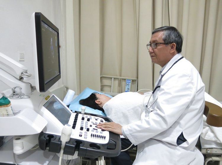
2.2 Echocardiography
The goal of echocardiography is to rule out extrinsic causes of acute right ventricular failure requiring urgent treatment, such as acute cardiac tamponade, and to assess right atrial pressure and arterial pressure. Pulmonary systolic, assess total right ventricular function. In addition, echocardiography is also used to assess ventricular wall tension (longitudinal contraction of the region and entire right ventricle).

This is an invasive diagnostic method, recommended for cases of acute right ventricular failure of unknown diagnosis or unresponsive to treatment. Pulmonary catheter hemodynamic assessment provides accurate and continuous information on left atrial, right atrial pressure, cardiac output, and pulmonary capillary resistance. However, this is an invasive method, so it is usually only indicated in some complicated cases.
3. Treatment of acute right ventricular failure
The treatment of acute right ventricular failure requires multi-specialty coordination. The treatment of the disease also varies according to the clinical signs: Treatment of the consequences of right ventricular failure, relief of symptoms (dyspnea, pain) and psychological treatment of the patient.
3.1 Treatment process for acute right ventricular failure
● Assess severity of the disease: Including clinical assessment (neurologic signs, arterial blood pressure, diuretics), imaging (echocardiogram, CT scan) ), evaluate biochemical tests (lactate, hepatic marquer, BNP, renal function, troponin) and invasive tests (central venous catheter, pulmonary artery catheter
● Identify factors that promote treatment: Sepsis, cardiac arrhythmia, drug discontinuation, At the same time, treat specific causes such as coronary intervention for right ventricular myocardial infarction, reperfusion for high-risk cases of pulmonary embolism
● Optimizing body fluids: Intravenous diuretics (if fluid overloaded), renal replacement (if patients are unresponsive to diuretics), careful fluid resuscitation if central venous pressure is low, avoid
● Maintain arterial blood pressure: Use Norepinephrine;
● Consider vasopressors to reduce filling pressure: Including Levosimendan, Dobutamine and CCBs phosphodiesterase inhibitor;
● Apply other methods of reducing afterload: inhaled prostacyclin, inhaled NO.
3.2 Perform treatment for acute right ventricular failure
● Optimizing fluid volume: The amount of fluid entering the patient's body can overload the right ventricle, increase cardiac wall tension, reduce constriction, and cause clearance The tricuspid valve is more severe, has an increased influence on the biventricular relationship, reduces left ventricular filling and, as a result, decreases cardiac output. Therefore, fluid should be introduced into the patient's body with caution under monitoring of central venous pressure (CVP) in case filling pressure is not increased and low arterial pressure is a contributing factor. right ventricular failure;
● Diuretics: When right ventricular failure worsens, diuretics are the first choice of treatment along with maintaining arterial pressure for most patients with concomitant signs of venous congestion. Diuretic therapy helps to redistribute fluid in the venous system, thereby rapidly improving clinical symptoms. The monitoring of urine output, adjustment of diuretics is carried out strictly according to the treatment regimen;
● Treatment of vasopressors and inotropes: Indicated in cases of right ventricular failure with hemodynamic disturbances. The use of these drugs should be strictly according to the treatment regimen. Some commonly prescribed drugs include:
○ Noradrenalin: Restore blood pressure, increase blood flow to the brain, coronary and other organs. This drug can improve systemic hemodynamics;
○ Dobutamin, Levosimendan and phosphodiesterase III inhibitors: Increase contractility and increase cardiac output. However, these drugs can aggravate hypotension and may need to be combined with noradrenalin.

● Mechanical circulatory support device:
○ Acute mechanical ventilation: Indicated in cases such as right ventricular infarction, acute pulmonary embolism, graft failure after heart transplant or after using assistive devices left ventricular assist;
Extracorporeal membrane oxygenation machine/Life Support Equipment (ECMO/ECLS): Provides oxygen quickly in a short time. However, after 5-10 days, ECMO should be changed or changed to intermediate or long-term devices to avoid complications due to ECMO such as infection, peri-catheter thrombosis, local infection or hypoperfusion of the extremities. ;
○ Right ventricular assist devices (RVADs): Can be inserted into the patient's body by percutaneous implantation or surgery, used for a long time (4 weeks). These devices can also coordinate with oxygen concentrators when needed. However, the use of a right ventricular assist device also carries a potential risk of bleeding complications or thrombosis;
● Heart transplant: As the last treatment option in cases of right ventricular failure that does not respond to the above treatments.
Acute right ventricular failure is a disease with a high degree of danger and relatively complicated treatment. Patients need early diagnosis and prompt medical treatment and intervention, to reduce complications and improve mortality
Please dial HOTLINE for more information or register for an appointment HERE. Download MyVinmec app to make appointments faster and to manage your bookings easily.


