This is an automatically translated article.
The article was professionally consulted by Specialist Doctor I Tran Cong Trinh - Radiologist - Radiology Department - Vinmec Central Park International General Hospital. The doctor has many years of experience in the field of diagnostic imaging.If not diagnosed and treated early, herniated disc can cause many dangerous complications, directly affecting the patient's ability to walk.
1. What is a herniated disc?
The spine has 23 discs and is located between the vertebrae with a double-sided convex lens (including annulus, mucinous nucleus, and cartilage plate). The disc has the elastic ability of the annulus and the physiological movement of the mucous nucleus, so it has high adaptability and elasticity, helping the spine to avoid strong shocks.Herniated disc occurs when these shocks happen too many times, the spine is damaged, the disc is compressed too much, causing the fibrous capsule to crack and the mucus nucleus to escape. Some causes of disc herniation are mainly due to:
Trauma: Acute traumatic factors and micro-traumatic factors are the primary cause of onset. Age: The older the age, the weaker the resistance of the annulus, the ability to synthesize Mucopolysaccharide and Collagen decreases, the chondrocyte cells lose their ability to self-regenerate. If the spine is subjected to a large load, trauma, or micro-trauma, it will cause a herniated disc. Occupation: Herniated disc often occurs in people who work in a restrained position, too hunchback or have limited mobility. Unhealthy habits: Sitting with your neck flexed, sleeping with too high a pillow, lifting heavy objects... causing disc damage. In addition, diet, pregnancy, obesity... are also factors that cause disc herniation. When there is a herniated disc, the patient will have symptoms such as:
Muscle weakness, numbness, tingling in one or both legs Dull pain sometimes intense, sitting or standing for too long also causes pain become intense. Pain in the lower back, spreading to the legs and buttocks; numbness, joint pain, limbs feeling weaker than usual Health severely reduced Mobility becomes difficult...

2. Diagnosis of disc herniation
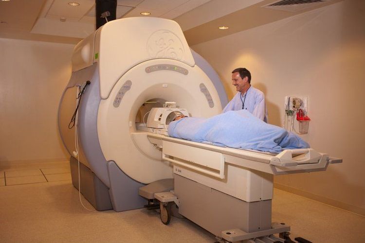
In addition to the above clinical symptoms, the following diagnoses are often used to confirm the disease:Routine X-ray: For X-rays of the spine, taken in 2 upright and inclined positions. Because the disc is a non-contrast organ, it is not possible to see the image directly, but to evaluate it indirectly through the change of the intervertebral space, the curve of the spine and adjacent vertebrae. Nerve root capsule: Contrast is injected into the subarachnoid space of the spinal cord. This is a method for indirect imaging of disc herniation with images of spinal stenosis, foramen synaptic, so it cannot distinguish compression caused by other causes. Computed tomography: This method has advantages in imaging the bone structure of the spine, is indicated in the diagnosis of spinal tuberculosis, spinal tumors... However, computed tomography is rarely used in Diagnosing disc herniation because the disc cannot be visualized directly. Magnetic resonance imaging: Magnetic resonance imaging is considered a "golden" test in the diagnosis of disc herniation, allowing the exclusion of lesions inside the spinal cord. Magnetic resonance imaging for canals, disc images with high resolution, observed in many different directions, is a safe and non-toxic method for patients. Therefore, magnetic resonance imaging is always the first choice in the diagnostic methods of disc herniation.

Please dial HOTLINE for more information or register for an appointment HERE. Download MyVinmec app to make appointments faster and to manage your bookings easily.


