This is an automatically translated article.
The article is professionally consulted by Master, Doctor Vu Quoc Anh - Department of Pediatrics - Neonatology - Vinmec Danang International General Hospital. Dr. Anh has nearly 10 years of experience as a resident doctor and treating doctor at Hue Central Hospital and Danang Children's Hospital.Urinary tract birth defects in children are relatively common. If not treated early, it can cause urinary tract infections, kidney failure and loss of function. The disease needs to be detected early for appropriate intervention.
1. Birth defects of genitourinary system
Urinary and genitourinary birth defects are defects in children, usually appearing right after birth and changing the shape and function of parts of this organ system. Genitourinary tract birth defects account for about one-third of all birth defects in humans.Birth defects can be in only one organ, or many organs are affected. It occurs in organs such as the kidneys, bladder, ureters, urethra, and the genitals in men are the penis, testes, and in girls, the vagina, ovaries, and uterus.
At present, the exact mechanism causing urogenital malformations in children is unknown. In some cases, parents also have similar foreign bodies or carry genes that cause birth defects and pass them on to their children.
Congenital urogenital malformations can lead to urinary tract infections, kidney damage, kidney failure, ... if not early intervention. Many malformations can be diagnosed with ultrasound. Immediately after birth, the defects will be diagnosed through postpartum screening, and tests to check the overall health of the newborn at the hospital.

2. Common urogenital birth defects
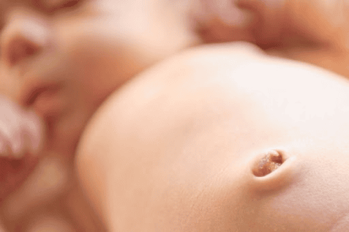
Congenital urinary tract malformations are relatively common in pediatric pathology. Some common birth defects include:Urinary tract stenosis: in a low deviation or narrowing of the urinary tract after circumcision. The patient has small urinary tract symptoms, difficulty urinating. Cure narrowing of the urinary opening by dilating and widening the urinary opening. Foreskin stenosis: symptoms of difficulty urinating, when urinating, the foreskin is bulging, the foreskin cannot be turned and the urinary opening is not visible. Treatment is by surgery, dilation or inversion. For the case of narrowing of the foreskin without the annulus, it is possible to gradually widen the foreskin or use a small pine to dilate, separate and clean the glans and glans. This method is simple, easy to do, does not cause pain for children and brings long-lasting results. In case of fibrous ring in the foreskin, the foreskin is blocked, surgery must be indicated. Urethral stricture: patients with difficulty urinating, small rays and urinary tract infections. Diagnose the location and extent of narrowing, narrow urethral length by urethrography. Depending on the length and severity of the narrowing, you can choose to dilate the urethra, cut open the narrow area by endoscopic surgery, or cut the narrow spot to reconnect the urethra or create a new urethral segment. Urethral diverticulum: usually occurs in boys, rarely in girls. The disease manifests itself right after birth. Children do not urinate, always pee. There is a fever due to a urinary tract infection. In the scrotum, there is usually a fairly round, stretchy mass, if pressed, urine will be released from the urinary hole. Urethrography to determine the position and size of the normal bag, Treatment with antibiotics and diverticulectomy, suture to reconstruct the urethra. Posterior urethral valve in boys: children have symptoms of difficulty urinating or continuous leakage of urine, so the bladder is often enlarged. Diagnosis is by X-ray or urethroscopy. The treatment method is endoscopic resection of the urethral valve. As for the urachal tube: see clear water seeping through the navel often or when the child urinates, the urine just comes out through the urinary opening at the top of the glans and just past the navel. Diagnosis is confirmed by cystography or by injecting methylene blue into the urethra. The disease usually resolves on its own with infants. If present, surgical removal of the urachal canal.

If you notice any unusual problems in your child, you should take your baby to see a doctor and consult a specialist.
>> See also: Birth defects from statistics to causes – Article written by Doctor Do Phuoc Huy, Vinmec High Technology Center
Please dial HOTLINE for more information or register for an appointment HERE. Download MyVinmec app to make appointments faster and to manage your bookings easily.


