This is an automatically translated article.
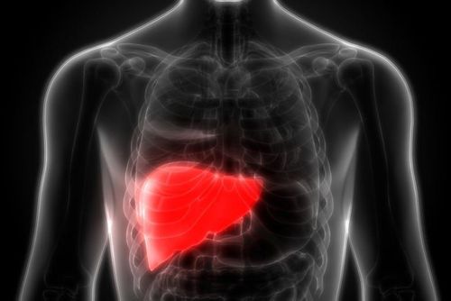
This article is professionally consulted by Master, Doctor Vu Huy Hoang - Radiologist - Department of Diagnostic Imaging and Nuclear Medicine - Vinmec Times City International Hospital.Computed tomography of the liver with biliary reconstruction is a modern imaging tool based on the classical CT liver technique. This is the preferred means in the diagnosis of hepatobiliary diseases in general and liver damage in particular. However, biliary reconstruction can also be performed by magnetic resonance imaging with higher resolution than computed tomography of the liver and promises to be more commonly indicated in the future.
1. Overview of liver computed tomography with biliary reconstruction
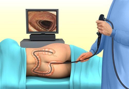
Along with the advancement of technology, computed tomography of the liver does not simply stop at examining the image and structure of the liver parenchyma in liver lesions but also allows the patient's biliary tract to be visualized. Helpful in the diagnosis of hepatobiliary diseases. The intrahepatic and extrahepatic biliary systems can both be examined and reconstructed with true structural images. This method has the potential to complement hepatobiliary ultrasound in cases of small, complicated lesions and to assess the overall structure of the liver and the associated biliary system, especially in obstructive pathologies. biliary tract such as cholangiocarcinoma, gallstones.Computed tomography of the liver with biliary reconstruction is a minimally invasive method in which the patient needs intravenous contrast injection. Therefore, the person being examined for imaging needs to be carefully examined about the history of drug allergies, especially to contrast agents, and carefully consulted and examined clinically before the procedure. In general, the method of computed tomography of the liver with biliary reconstruction is quite safe with a relatively low rate of complications during and after the technique. A few unwanted cases may come from the side effects of contrast agents. Image quality in computed tomography of the liver with biliary reconstruction depends on many factors such as imaging technique, exposure of the survey area, and the ability to maintain posture during the scan.

Trong chụp cắt lớp vi tính gan có dựng hình đường mật cần sử dụng thuốc cản quang
2. Indications and contraindications for computed tomography of the liver with biliary reconstruction
Cases that need to be performed computed tomography of the liver with biliary reconstruction include:Diagnosis of hepatobiliary diseases that abdominal ultrasound may miss or cannot evaluate in detail Abdominal ultrasound detects can detect localized liver lesions but of unknown nature Assess pressure on biliary system Monitor hepatobiliary system diseases when patients are difficult to detect by ultrasound such as overweight and obese people Hepatobiliary management should evaluate the vascular system and surrounding tissues such as primary hepatocellular carcinoma, malignancy from other organs with metastasis to the liver, and benign liver lesions. Obstructive diseases of the biliary system such as gallstones, cholangiocarcinoma The method of liver computed tomography with biliary reconstruction is quite safe, but not everyone can be indicated. Contraindications to computed tomography of the liver with biliary reconstruction include:
Contrast allergy Women suspected of being pregnant or are pregnant Chronic kidney diseases, renal failure

Bệnh nhân suy thận chống chỉ định thực hiện chụp cắt lớp vi tính gan
3. Steps to conduct computed tomography of the liver with biliary reconstruction
To ensure safety and quality of images obtained, computed tomography of the liver with biliary reconstruction needs to be performed sequentially according to the following steps:Preparation of instruments and facilities: the type of machine required is a scanner. Computerized tomography on 8 rows with necessary medical supplies such as syringes, needles, cotton, gauze, physiological saline, contrast agent, shockproof box. Prepare the patient: before the procedure, the patient should be carefully explained and advised about the procedure as well as the possible complications during and after the scan, and the patient should be advised to fast. at least 4 hours before. When entering the technique room, the patient needs to follow the instructions of the technician and the radiographer such as disassembling accessories and jewelry, changing uniforms, lying on his back, keeping the posture coordinated with the movements. correct breathing as required. In case the patient is too agitated or anxious, sedation can be prescribed at the discretion of the doctor.

Trong nhiều trường hợp, bệnh nhân được dùng thuốc an thần trước khi chụp cắt lớp vi tính
At Vinmec International General Hospital, the thoracic computed tomography scan is performed by a team of highly qualified and experienced doctors in the field of diagnostic imaging, along with equipment , modern machines for accurate and fast results.
Doctor Vu Huy Hoang has 10 years of experience working in the field of diagnostic imaging, formerly a Doctor at the Department of Diagnostic Imaging - Thai Nguyen Central Hospital. Currently, the doctor is working at the Department of Diagnostic Imaging - Vinmec Times City International Hospital.
To register for examination and treatment at Vinmec International General Hospital, you can contact the nationwide Vinmec Health System Hotline, or register online HERE