This is an automatically translated article.
Video content is professionally consulted by MSc, BS. Phung Thi Ly, Department of Obstetrics and Gynecology - Vinmec Times City International Hospital
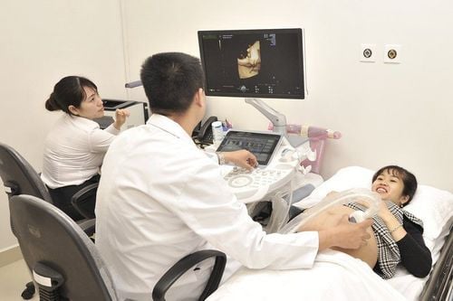
Ultrasound is a commonly used method of screening for fetal abnormalities, with high accuracy, helping pregnant women to detect fetal abnormalities early for early intervention. The following are common fetal abnormalities.
Cleft lip and cleft palate: 1 in 500 - 600 babies are born with this disease. There are many causes of the disease such as: genetics, environment, maternal medication or infection during pregnancy. This fetal malformation is usually detected at 21 to 24 weeks. Congenital heart defects: the abnormal formation and development of the heart and large blood vessels will cause congenital heart defects. About 1% of babies born alive may have heart defects. From 21 to 24 weeks is the golden time to help identify congenital heart defects most accurately. Down syndrome: due to an excess of chromosome 21, after birth 50% of children will have poor visual and hearing development. Pregnant women can check this fetal malformation most accurately from 12 to 14 weeks. Foot deformity: this is one of the most common fetal malformations, caused by the position of the fetus in the uterus. , the foot is compressed in the uterus (due to the large fetus, narrow mother's uterus, twins). This type of fetal malformation is usually detected during ultrasound at 12 to 14 weeks. In addition, the fetus may also experience some other malformations such as: skull malformations, abdominal wall cleft, hydrocephalus. hydrocephalus, spondylolisthesis, skeletal dysplasia, short limbs. Some birth defects can be treated in the womb, but some can only be treated once the baby is born.
Please dial HOTLINE for more information or register for an appointment HERE. Download MyVinmec app to make appointments faster and to manage your bookings easily.


