This is an automatically translated article.
Echocardiography in myocardial infarction by color Doppler method helps diagnose myocardial infarction when other diagnostic methods give unclear results, helping to assess the extent of damage to the myocardial infarction area. , prognosis and complications after myocardial infarction.
1. What is a heart attack?
Like any muscle tissue in the body, the heart needs a constant supply of blood and nutrients. If one of the major coronary arteries or small branches is suddenly blocked, the part of the heart that nourishes the blood vessels will be deprived of blood and oxygen, causing myocardial ischemia. If myocardial ischemia lasts too long, the heart muscle tissue will die causing angina, also known as myocardial infarction.
The cause of a heart attack is usually due to the atherosclerotic plaque on the vessel wall falling off, exposing the damaged blood vessel wall. Platelets in the blood will come to agglomerate at the injured site, forming a blood clot that blocks the blood vessel lumen. Common symptoms of a heart attack are angina pectoris, pain that varies from mild to severe, sharp like a stabbing knife, pain that can spread to the neck, lower jaw, shoulders, back, stomach,... Other symptoms include difficulty breathing, nausea, vomiting, sweating, dizziness, heart palpitations,...
Heart attack can cause sudden and rapid death. Patients may experience cardiac arrhythmias, cardiac arrest, coma, sudden death, etc. Therefore, early detection of myocardial infarction for timely treatment plays an extremely important role. Paraclinical methods to help evaluate myocardial infarction include: electrocardiogram, cardiac enzyme testing and cardiac biomarkers, echocardiography in myocardial infarction,... In which, Doppler echocardiography Coloring is a particularly valuable method, helping to diagnose myocardial infarction when other diagnostic methods give unclear results. , diagnosis of right ventricular infarction, assessment of mechanical complications and thrombosis,...

Người bị nhồi máu cơ tim có thể tử vong nhanh và đột ngột
2.Color Doppler echocardiography in the assessment of myocardial infarction
Doppler echocardiography is a non-invasive diagnostic technique that examines moving objects with a transducer that receives ultrasound waves. With the frequency signals emitted from the transducer of the ultrasound machine and the frequency received when surveying a moving object, the ultrasound machine will synthesize and display it on the screen in the form of colors, spectral waveforms or sound can be heard. Doppler ultrasound has the advantages of fast, high accuracy; Convenient when it can be done at the hospital bed, it plays an important role in detecting, classifying and treating emergencies.
See also: Doppler ultrasound to assess complications after myocardial infarction
2.1. Echocardiography in myocardial infarction with color Doppler Color Doppler ultrasound helps to confirm the diagnosis of acute myocardial infarction when the following phenomena are present:
There is regional movement disorder including reduced movement, inactivity (most common) ) or paradoxical movement. Cardiac wall thickness (diastolic): normal Healthy heart walls: compensatory increase in motion, but not in the presence of multivessel stenosis. Movement disorders often appear early. The sensitivity of Doppler echocardiography for transmural myocardial infarction is 88-100%, and for subendocardial infarction it is 86%. The specificity ranges from 53-94%. Ultrasound can predict the location of coronary artery damage with 90% sensitivity.
On the prognosis of myocardial infarction:
Regional motor index >2: severe prognosis (80%, 90%) Regional motor index >7: severe heart failure (88%, 57%) Mental function ventricular diastolic diastole, if E/A ratio >1, TG deceleration E (DT) ≤140ms, prognosis of death, rehospitalization, reconstruction and left ventricular dilatation after myocardial infarction. 2.2. Doppler ultrasound helps in differential diagnosis

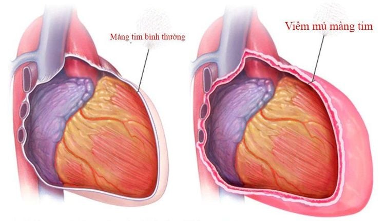
Siêu âm Doppler giúp chẩn đoán phân biệt nhồi máu cơ tim với bệnh viêm màng ngoài tim
Before a patient with persistent chest pain, echocardiography helps to differentiate myocardial infarction from other conditions such as:
Pericarditis Myocarditis Aortic dissection 2.3. Doppler ultrasound helps to detect complications after myocardial infarction 2.3.1. Complications of pericardial effusion Approximately 20-25% of patients with myocardial infarction have pericardial effusion on echocardiography after myocardial infarction. Doppler ultrasound helps to confirm the diagnosis, extent, and cause of pericardial effusion.
Pericardial effusion usually appears on 3-10 days, lasts 1 month. Common in patients with heart failure, transmural myocardial infarction, anterior wall.
2.3.2. Heart Aneurysm Complications Cardiac aneurysm occurs in 1⁄5 cases of myocardial infarction, this complication can appear early. The diagnostic criteria for cardiac aneurysm on echocardiography are:
Derived from myocardial infarction area Widespread neck of aneurysm, ventricular deformity during diastole, paradoxical movement during systole, thin heart wall 2.4. Complications of perforation of the interventricular septum after myocardial infarction

Sau nhồi máu cơ tim có thể gặp biến chứng thủng vách liên thất
Complications of perforation of the interventricular septum after myocardial infarction are rare, the rate is from 1-2%, but the equivalent is severe. When 2D ultrasound of the four chambers of the heart, the longitudinal axis to the nasopharynx can show a perforation in 60-80% of the cases, however, a perforation <5mm cannot be detected. If 2D ultrasound combined with color or contrast Doppler will give higher sensitivity and specificity.
2.5. Complications of mitral regurgitation after myocardial infarction Mitral regurgitation is a common complication after myocardial infarction, due to causes such as:
Dilation of the annulus secondary to left ventricular dilatation, papillary muscle dysfunction, ligament rupture , muscle papillae Doppler ultrasound helps identify and evaluate the extent of mitral regurgitation.
2.6. Left ventricular thrombosis complications Doppler ultrasound can definitively diagnose left ventricular thrombosis with a sensitivity of 77-95%, a specificity of 89-86%. In addition to assessing the size, location, shape, and mobility, ultrasound also helps to predict the risk of embolism and the progression of thrombosis.
Myocardial infarction causes many different complications, and Doppler ultrasound is the leading diagnostic method because of its simplicity, high accuracy, and safety for the patient. Therefore, when there are any warning signs of this condition, the patient should go to the doctor immediately so that the doctor can designate the appropriate diagnosis, thereby having the best treatment plan.
Currently, Vinmec International General Hospital is not only well-regarded for its modern facilities, experienced medical team, but also as an address for early examination and diagnosis. and successfully treated many cardiovascular diseases, including myocardial infarction. In addition, Vinmec is also equipped with a modern color Doppler ultrasound machine, giving clear image results for examination and diagnosis. After the results are available, the Department of Diagnostic Imaging also cooperates with many other specialties to determine the stage of the disease to have an effective treatment plan for myocardial infarction, early to bring good health to the patient.
Please dial HOTLINE for more information or register for an appointment HERE. Download MyVinmec app to make appointments faster and to manage your bookings easily.


