This is an automatically translated article.
Chiari malformation is classified into four types as I, II, II, IV. In particular, type I chiari malformation is common and milder than other types.
1. What is a chiari malformation?
Chiari malformation is a birth defect characterized by brain tissue extending into the spinal canal.
Chiari malformation occurs when part of the skull is distorted or abnormally small, putting pressure on the brain and compressing brain tissue downward. This malformation is usually associated with spinal hollow (or hydrocephalus), ventriculomegaly, or posterior fossa cranial stenosis.
The prevalence of Chiari malformation in the population is less than 1/1000. Most cases have no symptoms. The malformation is often discovered incidentally when the doctor assigns the patient to perform imaging techniques to diagnose other diseases.
2. Classification of Chiari . malformations
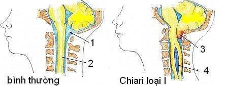
Dị dạng Chiari type 1
In 1891, based on the results of autopsies, the Austrian anatomist Hans Chiari (1851-1916) classified Chiari malformations into 4 types, type I, II, III, IV based on the severity of the disease. malformation.
Among them, type 1 chiari malformation is the most common and milder. Other types of malformations are often detected early, possibly right after birth because they can be accompanied by many other malformations such as meningeal herniation in the occipital cervical hinge region, gyrus malformation, and lower ventricle drop than normal. often,... This Chiari malformation classification system is still in use today.
2.1 Chiari malformation type 1 Chiari malformation type 1 occurs during fetal development, and is characterized by the position of the cerebellar amygdala moving more than 4mm below the occipital foramen into the cervical canal. Migration of the cerebellar amygdala can block the normal circulation of cerebrospinal fluid between the spinal canal and the intracranial space.
Chiari malformation can be associated with spinal hollow. About 30-50% of patients with type I chiari malformation have abnormalities in the base of the skull and spine such as:
Attachment of the first cervical vertebra to the base of the skull; partial adhesion of the first cervical vertebra to the second cervical vertebra; The upper part of the spine presses against the base of the skull, causing compression of the brain stem; Latent spina bifida or scoliosis The clinical symptoms of Chiari type 1 malformation vary slightly between children and adults, especially in neonates.
In adults, at the beginning, the disease often has no clinical symptoms. Only when there are signs of nerve compression can symptoms appear clearly such as: Visual and auditory disturbances, spinal cord dysfunction, headache, dizziness when changing positions, numbness of limbs. .
Signs of spinal cord compression, brain stem or cranial nerve pain also manifest as sleep apnea, limb tremors or glare, photophobia, limb weakness, muscle atrophy, hemispasm face,...

Người bị thoát vị tủy-màng tủy có nguy cơ dị dạng chiari loại II
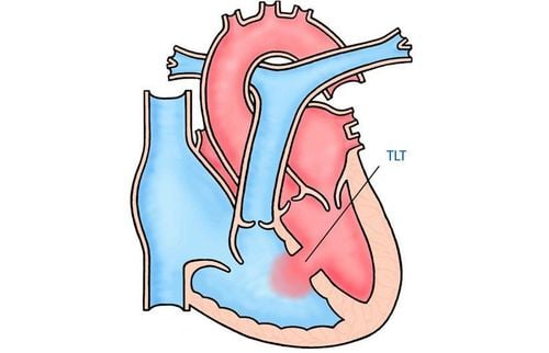
2.2 Arnold chiari malformation (type II chiari malformation) Arnold chiari malformation is used to refer to type II chiari malformation. This type of malformation occurs in patients with medullo-membranous hernias.
Hernia-meningeal hernia is a birth defect that occurs when the spinal cord and spinal column do not close properly during fetal development, the baby is born with part of the spinal cord that is open at the back.
Characteristic of Chiari malformation type II is that the patient has a neural tube defect, with downward migration of both the cerebellar pupa and medulla oblongata through the occipital foramen into the cervical spinal canal. The cerebellar amygdala is usually very low. Patient There are many other central malformations such as hydrocephalus, torsion of the medulla oblongata, hypoplasia of the cerebellar tent, hypoplasia of the corpus luteum,...
Patients with Arnold Chiari malformation will have symptoms. symptoms such as vomiting; breathing changes; arm weakness; downward movement of the eye, rapidly, involuntarily,...
2.3 Chiari malformation type III This type of Chiari malformation is very rare, it is a special form of posterior brain herniation. Patients present with a sac at the occipital neck joint containing the cerebellum and brain stem, often with hydrocephalus. Patients with this type of malformation often have an early death or severe psychosis if they survive.
2.3 Chiari malformation type IV This is the rarest and most severe type of Chiari malformation. The cerebellum is abnormally developed, with severe hypoplasia and aplasia of the cerebellum, along with many other serious malformations, more severe than children who die soon after birth.
3. How is Chiari malformation diagnosed and treated?
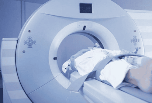
Chụp cắt lớp vi tính
To diagnose Chiari malformation, the doctor may assign the patient to perform a number of techniques such as:
Computed tomography (CT scan): Helps determine the size of the ventricles and detect blockages. Magnetic resonance imaging (MRI): Provides accurate images of the brain, cerebellum and spinal cord, thereby determining the degree of deformity and detecting the progression of the lesion. Myelogram: Helps to see the image of compression on the spinal cord or nerves due to deformity. Brain auditory evoked potential (BAER): Helps to check the integrity of the auditory conduction system, thereby determining whether the brain stem is working properly. Somatosensory evoked potential (SSEP): is a method of recording action potentials of nerves involved in sensation, thereby providing information about peripheral nerves, spinal cord and function brain function. Treatment for Chiari malformation depends on the type, severity, progression, and symptoms of the patient.
Please dial HOTLINE for more information or register for an appointment HERE. Download MyVinmec app to make appointments faster and to manage your bookings easily.
SEE MORE
Some common neonatal malformations that need early intervention Congenital urogenital malformations in children need early intervention Screening for birth defects