This is an automatically translated article.
Posted by Master, Doctor Lam Thi Kim Chi - Doctor of Radiology - Department of Diagnostic Imaging - Vinmec Danang International General Hospital
1. How to conduct an ultrasound of the spine?
Ultrasound transmits sound waves into the body through a hand-held device (transducer) capable of sending and receiving signals. The doctor will apply gel to the patient's back and place the probe on the area to be examined. The doctor will then analyze and evaluate the images recorded and displayed on the screen of the ultrasound machine. From there, diagnoses, methods of monitoring and treatment for each case are given. Ultrasound is usually quick, painless, non-radioactive, and harmless to the child.
Prepare the patient:
Usually does not need to prepare the baby before the ultrasound The baby should be dressed in clothing that exposes the back when asked. Babies can be fussy, making ultrasound difficult. Parents should prepare toys, bottles, movies... to attract the baby's attention.
2. Indications for pediatric spinal ultrasound
Indications for ultrasound of the spine of children in the following cases:
Screening for congenital marrow abnormalities. The child has an indentation or opening in the lower part of the spine. Newborns with congenital spondylolisthesis or spina bifida. Evaluation of the consequences of spinal cord injury during birth. In addition, some abnormal signs in the spine need to be screened as follows:
Sinus dermis. Localized hirsutism. Skin hemangioma or lipome in midline, midline. Anal malformation - rectum. Real estate dead end. Normal spinal ultrasound:
The spinal cord is a tubular, hypoechoic structure, clearly delimited by two hyperechoic margins. Inside the spinal cord there is an endothelial canal. The spinal cone is a tapered part of the spinal cord, usually located on the L1 - L2 lumbar spine. The terminal cord is less than 2mm thick. In addition, some other components in the spinal cord can also be evaluated such as the cauda equina, sacral sac, ...

3. Some malformations investigated on spinal ultrasound
3.1. Marrow adhesion syndrome
The spinal cone adheres to the lower part of the spine Often there are no clinical symptoms. Discovered only incidentally when examining the anorectal malformation system. Progression: dyskinesia, abnormal reflexes, sphincter disorder
3.2. Double spinal cord
There is a bifurcation of a cord or terminal cord. The two halves of the marrow are often disproportionate. Often with hair tufts or hyperpigmentation, spinal deformity.
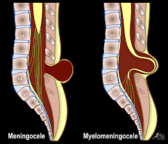
3.3. Meningeal hernia
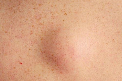
Herniation of the dura, arachnoid and cerebrospinal fluid in the subcutaneous sac of the dorsal region. It is common to see children with tumors in the sacrum, rising and falling, changing in size, increasing in the sitting position.

3.4. Sinus dermis
This is a thin tube surrounded by skin and connective tissue
Common in the sacroiliac area There is little hair, intermittent drainage Abscessization or inflammatory tissue mass in the sacrum.
3.5. Fat tumor
There is a round block of fairly solid density in the dead end area.
Spinal ultrasound is often done to evaluate the condition of the spinal cord in infants. This method is usually done when the child has abnormal signs in the sacrum, spine.
The Department of Diagnostic Imaging is one of the spearheads of Vinmec International General Hospital, equipped with the most modern and advanced equipment in the country as well as in the region. The department has 6 ultrasound rooms, 4 DR X-ray rooms (1 full-axis machine, 1 brightener, 1 synthesizer and 1 mammography machine), 2 DR mobile X-ray machines, 2 cutting rooms. multi-row computer class with receiver (1 128-series and 1 16-series), 2 Magnetic Resonance imaging rooms (1 3 Tesla machine and 1 1.5 Tesla machine), 1 interventional angiography room with 2 planes and 1 room measure bone mineral density. The doctors at the Faculty are well trained at home and abroad. The department also regularly cooperates with a team of experienced experts and doctors from the Department of Diagnostic Imaging, Bach Mai Hospital.
Please dial HOTLINE for more information or register for an appointment HERE. Download MyVinmec app to make appointments faster and to manage your bookings easily.


