This is an automatically translated article.
The article was written by Master - Doctor Mai Vien Phuong - Head of Gastrointestinal Endoscopy Unit - Department of Medical Examination and Internal Medicine - Vinmec Central Park International General Hospital.Gastrointestinal fistula after surgery is a very big problem, very complicated. It's big because it can be encountered everywhere, at any time, in any situation. Complicated by the clinical manifestations are very variable, can be very mild but can also be very severe. Prognosis varies widely, depending on the lesion, care, and treatment. All gastrointestinal surgeons can experience this complication, with varying rates.
1. The term “Enterocutaneous fistula”
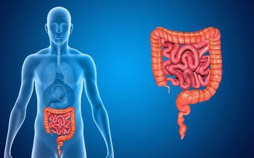
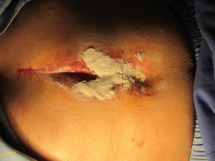
The term "Enterocutaneous fistula" refers to a lesion of the gastrointestinal tract, through which the contents of the tube pass through the chest wall, abdominal wall, ... and out of the body. Gastrointestinal fistula does not include lesions in which the contents of the gastrointestinal tract through the fistula at the suture line, the rupture at the junction, the perforation, the tear, the necrosis in the intestinal wall, etc., into the abdomen causing peritonitis . Lesions through which the contents of the gastrointestinal tract in one place enter another part of the gastrointestinal tract are called "internal fistulas". Another concept is “anal fistula lesions”, which begin with an infection in the Morgagni valves of the anus. On this infected lesion an abscess forms. Abscesses break down the layers of the wall of the rectum and anal canal to drain the pus into the anal canal or out, around the anal opening.
Rò tiêu hóa còn gọi là thương tổn rò hậu môn
2. Digestive fluid includes what?
Digestive fluid is secreted from 4 types of glands of the alimentary canal, with the daily amount as follows:Saliva from the salivary glands : 800 - 1500 ml. Gastric secretions from the glands of the stomach: 2000. Bile from the glands of the liver: 1000. Pancreatic juices from the secretions of the pancreas: 1200 - 1500. In total per day, the amount of fluid secreted into the gastrointestinal tract is about 5-6 liters. Saliva: Made of 3 pairs of glands: parotid, submandibular, and sublingual. In addition, glands in the mouth and tongue are involved. Saliva contains amylase, mucus and some electrolytes. Saliva is alkaline, favorable for amylase activity, which helps prevent tooth decay by neutralizing bacterial acids.
Gastric juice: Secreted by 3 types of cervical cells, main cells and parietal cells.
Glandular cells secrete mucus. Mucus is responsible for protecting the stomach lining, avoiding the effects of pepsin. Main cells secrete pepsinogen. This substance is a precursor to pepsin. Parietal cells secrete hydrochloric acid, which converts pepsinogen into pepsin. Pepsin has the effect of causing gastric and duodenal ulcers. Bile: Made from liver cells. Once secreted, the bile flows into the intrahepatic bile ducts, followed by the extrahepatic bile ducts, and is stored in the gallbladder. During digestion, the gallbladder contracts, expels bile into the common bile duct, and then empties into the duodenum. In bile there are bile salts, bilirubin, cholesterol, lecithin, ions and water. Bile is essential for fat absorption, secretin stimulates bile secretion. Nerve X and cholecystokinin increase the contraction of the gallbladder, the total amount of bile from the gallbladder through the common bile duct, the ampulla of Vater, the sphincter of Oddi into the duodenum. Pancreatic juice: The pancreas has 3 types of cells: endocrine cells, exocrine cells, and duct cells. Endocrine cells secrete insulin. Exocrine cells secrete enzymes: protease, lipase, amylase, nuclease. Tubular cells excrete bicarbonate solutions. The substances acetylcholine, gastrin, cholecystokinin, secretin have the effect of regulating pancreatic secretion. In addition to the glands that secrete digestive juices mentioned above, there are also glands of the small intestine. The Brunner gland is located between the pylorus and the ball of Vater. This gland secretes mucus. The gland is active when the mucosa is stimulated by the X nerve or secretin. The gland is secreted by the sympathetic nervous system. Lieberkuhn glands are present throughout the mesentery of the small intestine, secreting an extracellular fluid-like fluid of about 1800 ml per day. This fluid is rapidly absorbed by the intestinal villi.
3. Causes of gastrointestinal leakage after surgery
Swollen stitches: Any stitch, a seam is likely to leak, poop. Errors in stitching technique, defects in equipment, bad needles can cause the stitches to leak at any time. This is often a threat to the surgeon.Blinking at the joint: Swollen and broken joints occur more often than at stitches. The cause of anastomosis may be due to:
Nutritional anemia. When the intestines are twisted, the blood vessels are blocked. When making a mouthpiece, cut too much of the mesentery. When the mouth floats, the intestinal loop is stretched. Anastomosis on the segment of intestine with diseased tissue. Abdominal cavity with turbid, purulent, fecal matter. Large bowel lumen during emergency colon surgery or elective surgery where the bowel is not cleaned. The patient's condition is not good: Asthenia, malnutrition, chronic disease, cancer,...
Rò tiêu hóa do xì bục miệng nối
4. Gastrointestinal fistula lesions are common in what organs?
Esophagus Esophageal injuries are uncommon. The most common is leaky mouth after esophagectomy due to esophageal cancer, cardiatric cancer, after esophagectomy due to burns. The reason is that the esophagus is not well supplied with blood compared to other parts of the digestive tract. According to doctors Nguyen Anh Tuan and Pham Duy Hien at 108 Military Hospital, there were 2 patients with oesophageal-intestinal fistula in 149 cases of total gastrectomy due to cancer. In these 2 patients with anastomosis, 1 was sutured, the fistula led to an abscess, the abscess was drained, and both were discharged from the hospital in stable condition. Similarly, Dr. Pham Huu Thien Chi at Cho Ray hospital has 46 patients with cancer of the heart and the lower third of the esophagus treated by esophagectomy through the diaphragmatic slit with 4 leaks, the rate is 8.68 %.Stomach: The stomach is less prone to bloating because there is a rich system of nourishing blood vessels. After temporary gastric bypass surgery, Fontan bag type, after conventional extubation, this stoma closes on its own. However, many times, it does not close itself, but creates a leak.
Duodenal: Fissure at the duodenal apex occurred after Billroth II gastrectomy. This lesion a few decades ago appeared quite a lot, but today it has been and is reduced completely due to the results of medical treatment, peptic ulcer is greatly reduced. If there is, the damage is not too big, not as callous as before. The ileum: Injuries or abdominal wounds, if there is damage to the intestine, will cause peritonitis, not fistula, because the jejunum is completely located in the peritoneal cavity and is easily mobile. The cause of jejunal fistula is mainly due to suture leak or anastomosis. The last 20cm of the ileum is not well nourished and has only one vascular arch (arch of Trèves).
Colorectal: The anastomosis of the ileum-colon, colon-colon-rectum is at high risk of perforation, especially during emergency surgery or during elective surgery where the colon is not cleaned before surgery. There is still a lot of stool or water in the colon. Because colorectal fistula does not cause dehydration, it does not affect health, does not make patients uncomfortable, does not affect activities much, sometimes the disease lasts for many years.
Please dial HOTLINE for more information or register for an appointment HERE. Download MyVinmec app to make appointments faster and to manage your bookings easily.
ReferencesNguyen Dinh Hoi. Gastrointestinal fistula, Gastrointestinal surgery. Medical Publishing House 1994, 225-253. Nguyen Hoang Bac. Colonic lavage during surgery. Thesis of Master of Science in Medicine and Pharmacy, University of Medicine and Pharmacy, Ho Chi Minh City 1997. Nguyen Trung Tin. Duodenal fistula after traumatic surgery and duodenal wound: clinical features and management attitude, Practical Medicine 2001, 401, 8:15-18.


