This is an automatically translated article.
The article was written by Master, Doctor Mai Xuan Thien - Emergency Doctor - Emergency Department - Vinmec Times City International General HospitalCarotid artery dissection is common in young people; The disease has signs and symptoms similar to other diseases, so it is difficult to diagnose the disease at the first time when the patient comes to the doctor. Therefore, accurate diagnosis of the disease plays an important role in helping patients reduce mortality and sequelae of the disease.
1. What is carotid artery dissection?
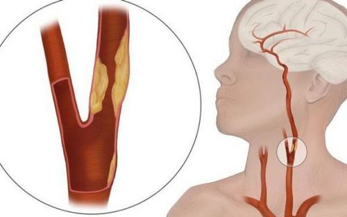
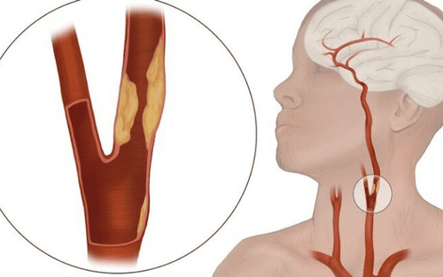
Carotid dissection is when the layers of the carotid artery wall separate. This damage affects cerebral blood flow and can lead to stroke. Furthermore, it can occur in the intracranial or extracranial segments, causing subarachnoid hemorrhage or presenting as ischemic stroke.2. Causes of disease
Carotid dissection usually occurs spontaneously when there is tearing of the intima of the carotid artery, creating a hematoma in the vessel wall. This tear can be spontaneous or traumatic. A hematoma in the vessel wall causes narrowing and results in the formation of a blood clot in the vessel wall.Traumatic dissection can be caused by blunt trauma or by oblique trauma. Concussion injuries can be severe (such as a car accident, ...) or mild (such as from exercises that impact the cervical spine,...). Traffic accidents when the vehicle is decelerating rapidly can cause the neck to be excessively flexed or rotated, leading to tearing of the inner lining of the carotid artery.
Spontaneous carotid dissection usually occurs in cases where there is a family history of dissection. Marfan syndrome, Ehlers-Danlos syndrome, fibromuscular dysplasia, and other connective tissue disorders increase the risk of this disease. The long styloid in Eagle syndrome can also cause spontaneous carotid dissection.
3. Epidemiology
Carotid artery dissection occurs in all age groups. They account for 2.5% of all strokes. It is the most common cause of stroke in people under the age of 40. In all young adults, 20% of accidental cerebrovascular injuries result in aortic dissection. The mean age of the disease is 40, and it seems that the prevalence is slightly higher in men than in women
4. Pathophysiology
The endothelium of the arterial wall is suddenly torn by the factors listed above, causing blood flow to flow into the inner layer of the artery wall, which is favorable for the formation of a hematoma there and the formation of blood clots. prosthetic lumen in the arterial wall.When blood enters the false lumen, it narrows and can lead to complete stenosis of the carotid artery. This is a dynamic process that can narrow the lumen or dilate the artery wall, depending on whether the hematoma grows inward or outward of the lumen.
This process leads to a stroke if it completely occludes the carotid artery. It may also be the site of thrombus formation that can cause dissection in other segments, possibly causing transient ischemic stroke. If the blood vessel ruptures into the intracranial segment, it may cause subarachnoid hemorrhage or may form a pseudoaneurysm.
5. Clinical symptoms
There are many different clinical manifestations of carotid dissection, so the diagnosis is a real challenge for the clinician.Symptoms of the disease can range from no symptoms to an acute stroke. The classic symptoms are described as: Headache, eye pain, face pain and neck pain. Pain is usually unilateral.
Horner's syndrome may present if a hematoma in the neck causes compression of the cervical sympathetic plexus. When symptoms such as stroke present with these symptoms can make diagnosis less difficult. A family history of carotid dissection or a connective tissue disorder may be an indication of a higher incidence.
Circumstances in which the disease occurs (after trauma) may suggest a clearer diagnosis, but mild trauma (such as cervical spine therapies in therapy) will also make it more difficult . Unfortunately, in many cases there is neither pain nor obvious mechanism of injury that makes a correct diagnosis difficult.
6. Diagnosis
A thorough family history can aid in the diagnosis. In addition, many tools can help confirm the diagnosis such as:Carotid ultrasound: Minimal, minimally invasive method Computed tomography angiography (CTA): Allows visualization of arterial segments in skull. CT of the brain to identify acute stroke or intracranial bleeding. Cranial magnetic resonance imaging (MRI) : Can be substituted if the patient has a contraindication to CTA but is less sensitive.

7. Treatment
Treatment of carotid dissection depends on many factors, such as: whether the cause is traumatic or spontaneous, and whether the patient presents with a stroke. The location of the intracranial or extracranial dissection also changes the treatment plan. Active bleeding associated with hematoma extension is also a factor in determining treatment.If there are no contraindications, antiplatelet agents, or systemic anticoagulants, can be used to reduce the risk of stroke. Carotid stenting may be warranted, especially if anticoagulation is contraindicated or if medical therapy fails, the 1-year recurrence rate is 0-10%
Carotid artery dissection with May cause mild, moderate symptoms, or severe neurological deficits and even death. The prognosis of the disease is very variable depending on the time of diagnosis, whether there has been a stroke event. All patients are at very high risk of ischemic stroke, and intracranial bleeding and specific anticoagulation therapy also increase this risk.
This is a rare disease and very difficult to diagnose. Symptoms range from mild to life-threatening. The goal of treatment is to reduce the risk of stroke and worsen symptoms.
To minimize complications as well as detect the disease in time, if the patient shows clinical symptoms, it is best to go to reputable medical facilities for screening and diagnosis. Currently, at Vinmec International General Hospital, Doppler ultrasound technology is used to evaluate blood flow through the carotid artery. At the same time, combined with the most modern generations of color ultrasound machines in carotid examination and treatment such as GE Healthcarecar's Logig E9 ultrasound machine with full options, the transducers have HD resolution for images. Clear, accurate assessment of damage.
Besides, at Vinmec, there is a full team of experienced radiologists and doctors who will greatly assist in diagnosing and detecting abnormal signs early in order to come up with a method. The most accurate and timely treatment, improving the effectiveness of the treatment process and minimizing complications.
Please dial HOTLINE for more information or register for an appointment HERE. Download MyVinmec app to make appointments faster and to manage your bookings easily.


