This is an automatically translated article.
The article was professionally consulted by Specialist Doctor I Vu Thi Hanh - Department of Diagnostic Imaging - Vinmec Hai Phong International General Hospital.Ultrasound cannot completely differentiate thyroid cancer from other thyroid conditions. This is because ultrasound cannot visualize at the cellular level like pathology. To accurately diagnose the disease, it is necessary to combine different tests.
1. Ultrasound in thyroid cancer diagnosis
Can ultrasound detect thyroid cancer? Ultrasound is not usually used as a diagnostic tool to confirm thyroid cancer, but needs to be combined with other tests such as biopsy, fine needle aspiration cytology. It only helps to determine the abnormal nature of the tumor to predict and give suggestions for accurate diagnosis of the disease. Specifically, the role of thyroid ultrasound is described as follows:Classifying the tumor (solid, cystic, mixed)
Assessing adjacent structures
Determining the location of the tumor within or outside the thyroid gland
● Determining the cause of hyperthyroidism
● Determining the cause of hypothyroidism
● Postoperative complications such as abscess, edema
Multinodular goiter (MNG): Monitor nodules Abnormalities in the thyroid gland
Support injection, suction or biopsy
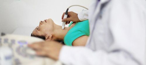
Siêu âm tuyến giáp giúp xác định nguyên nhân gây cường giáp
Fat roll in the corner of the neck
Lymphadenitis
● Cleft cyst (ultrasound on one side of the thyroid gland, faintly visible. via)
Thyroid cyst (visible in the middle of the thyroid on ultrasound)
Parathyroid mass (usually small and visible on thyroid ultrasound)
2. Limitations of ultrasound in thyroid cancer diagnosis
Ultrasound is subjective in nature, so the results of ultrasound depend a lot on the expertise of each doctor.3. Thyroid ultrasound procedure
3.1 Preparation
Collarless tops ● Remove jewelry around the neckShoulder / chest scarf
● Place pillows or towels under the shoulders (if needed) (Put pillows and cushions under the shoulders to raise the neck).
● When examining, it is necessary to perform some swallowing manipulations, turn the neck, do the Valsava maneuver to determine the origin of the lesion as well as distinguish it from neighboring lesions,
3.2 Technical equipment
Linear probe 7.5-10MHz. ● Use a machine capable of Doppler examination to assess the perfusion of lesions in the thyroid gland and thyroid gland.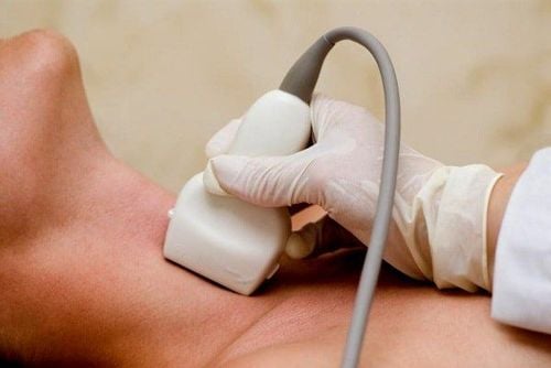
Thiết bị siêu âm Doppler giúp quan sát rõ các mạch máu xung quanh tuyến giáp
3.3 Conducting ultrasound
The sonographer will begin to place the transducer on the neck area, scan across the midline to evaluate tracheal deviation and differential diagnosis with some obvious pathologies Tilt the patient's head to the opposite side and scan down horizontally (scanning in the transverse section from top to bottom with the examination axis being the carotid bundle).● Rotate the transducer vertically and scan from the mid-to-side area
● Repeat these operations on the other side by tilting the head to the doctor's side
● The patient moves to a straight, scanning head and neck position transducers vertically and horizontally
Scan down each side of the neck horizontally for signs of differential diagnosis
4. Types of thyroid cancer
Thyroid cancer occurs in about 1% of thyroid nodules. Papillary carcinoma is the most common malignancy. The incidence of chemoradiocarcinoma is significantly increased in patients receiving radiation therapy to the head/neck region.Papillary carcinoma 78%
Follicular carcinoma 17%
Medullary carcinoma 4%
Cell carcinoma 1%
Thyroid lymphoma - rare
Cancer thyroid metastases - rare
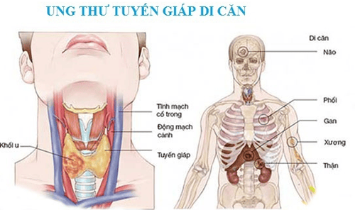
Ung thư tuyến giáp di căn là một ung thư hiếm gặp
5. Ultrasound in the diagnosis of malignant thyroid cancer
Ultrasound image of malignant thyroid cancer is shown through thyroid nodules with the following properties:● Hypoechoic mass
● Irregular mass border.
● The long axis is perpendicular to the skin surface.
Increase angiogenesis in the mass.
● Microcalcifications (but not large or peripheral calcifications)
Cervical lymphadenopathy (circular lymph nodes, loss of lymph node structure).
6. Some other tests in the diagnosis of thyroid cancer
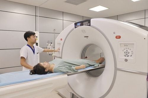
Fine-needle aspiration thyroid cytology / Ultrasound-guided needle biopsy. Biopsy : A method of using a fine needle or needle to take cells from the thyroid nucleus to send the pathology to read the results.● CT scan
CT is a method in addition to determining the structure and diagnosis of thyroid tumors, it can assess the stage of the disease (evaluate invasion, metastasis ..).
● PET scan
The PET test uses a small amount of radioactive material, a PET scanner, and a connected computer to look at body tissues. PET scans help see changes at the cellular level that help detect early-stage cancers and check how far they've spread.

Chụp CT giúp chẩn đoán ung thư tuyến giáp
● Genetic testing
Based on your family history, your doctor may recommend genetic testing to look for abnormal genes that cause cancer. This is the basis for suspecting thyroid cancer and other cancers.
Most thyroid diseases are ignored because the symptoms of the disease are often unclear, easily confused with many other diseases (except for large thyroid tumors causing protrusion of the neck, most of the adenomas are thyroid is ignored because there are no symptoms). When the disease has obvious symptoms, it is often at a dangerous stage, especially thyroid cancer (retired). Therefore, Vinmec International General Hospital has now been implementing a screening package for thyroid diseases. Vinmec's screening and screening package for thyroid diseases helps:
● Check thyroid function.
● Screening & early detection of common thyroid diseases such as simple goiter, hyperthyroidism, hypothyroidism, thyroiditis, thyroid nodules, thyroid cancer (especially thyroid cancer)
● If the client has thyroid tumors that are suspected of malignancy, thyroid cytology or thyroid biopsy will be performed to confirm the diagnosis.
When registering for the package of screening and screening for thyroid diseases, customers will be examined and consulted with an Endocrinologist; Thyroid ultrasound; Screening tests for thyroid diseases: FT3, FT4, TSH, Anti – TPO, Anti TG; With the most modern imaging methods, thyroid cancer screening can be detected since the patient has no symptoms.
Doctor Hanh has 12 years of experience in the field of Diagnostic Imaging. The doctor has the strength to train & perform tissue elastography with compression pulse and doppler echocardiography, cell aspiration, thyroid cyst and mammary gland under the guidance of ultrasound.
For advice on Thyroid Disease Screening Package, as well as other service packages at Vinmec International General Hospital, you can contact HERE.
References: webmd.com, ultrasoundpaedia.com