This is an automatically translated article.
The article is professionally consulted by Master, Doctor Ton Nu Tra My - Department of Diagnostic Imaging - Vinmec Central Park International General Hospital.Fibrocystic breast disease is a fairly common disease in young women of childbearing age. Fibrocystic breast tumors are mostly benign, but a small percentage can progress to cancer. Therefore, early diagnosis and follow-up play an important role in the treatment of the disease.
1. What is a fibroadenoma?
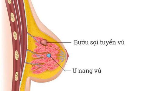
Fibrocystomas are a benign breast tumor that accounts for about 25% of all breast pathologies. Fibroids are solid, noncancerous tumors of the breast that usually occur in women between the ages of 15 and 35. . Can be detected incidentally, observed or palpable. Tumors are characterized by:Smooth, smooth, round or long. Touching shows that the tumor is hard, clearly bordered, when gently pressed, the tumor can move slightly, not sticking to other tissues and glands. Usually painless, it may feel like a marble in the breast, which can easily move under the skin on examination. Fibroadenomas vary in size and they can either enlarge or shrink on their own. You may have one or more fibroids in one or both breasts. The disease often appears with manifestations such as palpable lumps and lumps in the breast, a slight pain, the phenomenon is more pronounced when the menstrual cycle is approaching. Fibrocystic breast disease is a benign disease, rarely turning into cancer. However, the tumor still has a rate of turning into a malignant tumor, so the patient should not be subjective.
Fibrocystic breast tumors are less common in postmenopausal women, in cases where fibroadenomas have been present, the tumor tends to shrink as women begin menopause.
If the disease is not monitored and treated, it can lead to some of the following consequences:
If it is a malignant tumor, the patient may be detected at a late stage, affecting the treatment results. Large tumors can cause patients many uncomfortable feelings, pain, loss of aesthetics affecting life. Large tumors make surgery difficult and risk leaving bad scars for the patient. Causing anxiety, stress and guilt for patients

2. Can ultrasound detect fibroadenomas in the breast?
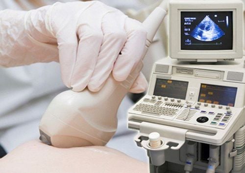
Breast ultrasound uses sound waves to create pictures of the inside of the breast. A breast ultrasound can help your doctor determine if a lump in your breast is solid or filled with fluid. A solid mass is more likely to be a fibroid; a fluid-filled mass is most likely a cyst.Breast ultrasound is indicated for any lump in the breast before other diagnostic tests are performed. Breast ultrasound helps assess the location, size, number, and nature of the tumor for the initial diagnosis. Therefore, breast ultrasound cannot accurately tell whether it is a fibroadenoma or not, but only allows suspicion and needs to conduct other tests such as:
Mammogram to evaluate the tumor in the breast. A mammogram (also called a mammogram) is a technique that uses X-rays to create images of the breast. A fibroadenoma on mammogram is usually a mass with well-defined margins, with even margins. Fine-needle aspiration cytology (FNA): is a technique that uses a small needle under ultrasound guidance, pierces the breast tumor, creates negative pressure and withdraws the contents contained within the tumor for cytology examination. cytology. This helps to suggest whether it is a fibroadenoma or not. Core Needle Biopsy (CNB): under ultrasound guidance is a technique that uses a needle with a core larger than that of fine needle aspiration cytology, under ultrasound guidance to take tissue samples from a tumor for tissue testing. pathology. From there, it helps to confirm whether the breast tumor is a fibroadenoma or not. Breast biopsy under the guidance of X-rays (Stereotaxic): is a technique of taking breast tissue samples under the 3-dimensional positioning of a mammogram machine for histopathological examination. Therefore, it helps to know whether the breast tumor is a fibroadenoma or not. MRI-guided biopsy (MRI-guided biopsy): is a technique to take a breast tissue sample under the 3-dimensional positioning of the breast magnetic resonance machine to do histopathological examination and know the nature of the breast tumor. fibroadenoma or not.

3. What to do with fibroadenoma?
If the tumor is less than 2cm in size, the patient only needs to continue to monitor the tumor status, no need to intervene with other solutions if the tumor does not cause much discomfort or troublePatients need periodic monitoring every 6 months to monitor and promptly detect abnormal changes of the tumor, detect early if the tumor changes into a malignant tumor or rapidly increases in size, affecting the patient's aesthetics.
If the size of the tumor is large, the patient is often indicated to remove the entire tumor.
At Vinmec International General Hospital, there is a Breast Cancer Screening Package to help detect breast cancer early even when there are no symptoms.
Breast cancer screening package at Vinmec for the following subjects:
Female customers, over 40 years old. Customers wishing to be able to screen for breast cancer Customers are at high risk of cancer – especially customers with a family history of breast cancer. Women of reproductive age, perimenopause and menopause. Women who are having symptoms of breast cancer such as: pain in the breast, lump in the breast, etc
Please dial HOTLINE for more information or register for an appointment HERE. Download MyVinmec app to make appointments faster and to manage your bookings easily.
SEE MOREBenign fibroadenoma need surgery? Treatment of benign fibroadenomas of the breast Learn about giant fibroadenomas of the breast


