This is an automatically translated article.
The article was professionally consulted by Specialist Doctor II Do Tuong Huan - Oncologist - Oncology Center - Vinmec Central Park International General Hospital.In Vietnam, lung cancer is one of the most common cancers, difficult to treat and causes death with a high rate, only after liver cancer. Currently, bronchoscopy is being applied to detect and treat lung cancer in time.
1. What is bronchoscopy?
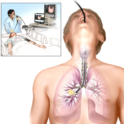
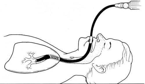
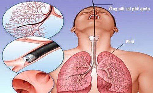
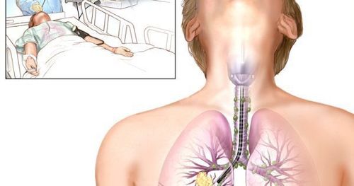
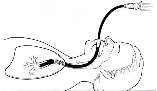
Bronchoscopy is a technique used in medicine, performed by passing a bronchoscope through the nose or mouth of the patient into the trachea or down the airways, penetrating deep inside the lungs to Observe the lesions there. In addition, bronchoscopy can take samples for testing, biopsy or treatment of lesions.Before appointing bronchoscopy, the doctor will conduct a comprehensive examination to see if the patient's condition is suitable for performing bronchoscopy. The patient is also responsible for informing the doctor about the medical condition related to the heart, hematology as well as the status of drug use in treatment, because some drugs can increase the risk of bleeding during treatment. endoscopic procedures such as anticoagulants, pain relievers (aspirin, ibuprofen...).
Before bronchoscopy, patients will be injected with drugs to reduce cough and relax, numb the nose and throat, measure the cardiovascular index and blood pressure. During the endoscopy, the patient will be connected to a machine that monitors the blood oxygen status and pulse. Note that the patient needs to fast for at least 4 hours before the endoscopy procedure to avoid reflux of food into the respiratory tract.
After the end of endoscopy, the patient continues to be monitored for another time, after 2-3 hours, if drinking water does not choke, then he can eat and drink normally. Patients may cough up bloody sputum 2-3 days after endoscopy and this is completely normal.
Bronchoscopy can be considered a relatively safe diagnostic procedure, however, there may still be some mild complications after performing such as: Shock due to hypervagal reaction, pneumothorax, coughing up blood, fever after endoscopy....
Nội soi phế quản có thể coi là một thủ thuật chẩn đoán tương đối an toàn
2. Bronchoscopy in the diagnosis of lung cancer
Lung cancer is currently one of the cancers with the highest mortality rate today, either in Vietnam or in the world. Most patients are often diagnosed with late-stage lung cancer and have a poor prognosis.Currently, the sputum test for cancer cells has a relatively low sensitivity due to errors in sample collection or preparation, while bronchoscopy is the current leading procedure in the early diagnosis of lung cancer. , or pre-invasive dysplastic lesions.
Diagnostic techniques used in bronchoscopy
Flexible bronchoscopy with white light or bronchoscopy with video: Support to clearly observe the central airway, but low sensitivity. Autofluorescence bronchoscopy (AFB): Assists in detecting pre-invasive endobronchial lesions. However, recent studies have shown that areas of abnormal fluorescence but benign histology, chromosomal changes are a sign of increased cancer index. High-magnification bronchoscopy: Performing bronchoscopy in combination with video allows magnifying the bronchi to 100-110 times, thereby allowing a clearer view of the bronchial mucosal microvasculature. , supporting the detection of dysplasia or early cancer. Narrow band imaging (NBI) : Supports a clearer view of the submucosal vascular network instead of using wideband in standard video. This technique is capable of detecting increased angiogenesis, the abnormal twisting of the vascular network during cancer production. Optical tomography (OCT): Produces high-resolution images of subsurface structures like ultrasound, but uses near-infrared light instead of ultrasound. OCT can acquire images of cells and extracellular areas by analyzing scattering with spatial resolution from 3–15 m wide and 2 mm deep, thereby providing images that are close to histology. in the bronchial wall. In addition, OCT also helps distinguish dysplasia from metaplasia, hyperplasia from normal tissue, lung cancer from invasive cancer. The severity of the histopathological stage is proportional to the gradual increase in epithelial thickness. In case the cell nucleus is darker and less bright, the basement membrane is interrupted or lost, which is a sign of invasive cancer.
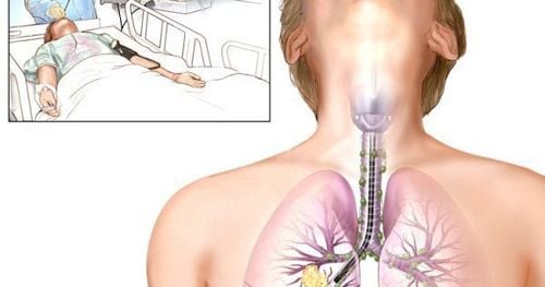
Nộii soi phế quản có thể cho lấy mẫu bệnh phẩm làm xét nghiệm
3. Bronchoscopy in the treatment of lung cancer
Currently, surgery is still considered the gold standard in the treatment of lung cancer with an 80-90% 5-year survival rate. However, its disadvantage is the removal of normal lung tissue in some cases.Bronchoscopy is an invasive technique to treat lung cancer with less health effects, lower costs than surgery with similar results, can be used to treat lung cancer in some cases where indicated.
Some lung cancer treatments through bronchoscopy
Endobronchial ablation: When electric current is introduced into the target tissue by rods or noose, heat is generated. Argon Plasma Coagulation - APC: Produces a plasma stream, through which uniform energy is transferred to and destroys the tumor. APC is effective for small and medium-sized tissues, but less effective for large tumors. Cryotherapy: Induces cell death due to rapid repeated freezing and slow thawing. This method works well with water-bearing tissue, less effective with less vascular tissue such as cartilage and connective tissue. Laser: Using laser is suitable for bronchoscopy due to its high energy. The effectiveness of the laser depends on the wave length. Photodynamic therapy (PDT): Kills lung cancer cells through the interaction between tumor-selective photosensitizers and laser light. The combination of light-sensitive molecules of particular wavelengths and tissue oxygen produces activated oxygen species capable of causing necrosis of cancer cells. Brachytherapy: This is done by placing a radioactive source at the tumor site. The advantage of this method is its high speed, thereby avoiding damage to healthy tissue compared to external radiation therapy. In the absence of lymph node metastases, internal radiation therapy in patients with lung cancer may yield positive results. The choice of technique for diagnosing and treating lung cancer through bronchoscopy depends on the body characteristics, disease status of each patient, and the medical equipment of the treatment unit. The treating physician needs to carefully examine, depending on the condition to apply the best method in diagnosis and treatment.
Flexible bronchoscopy technique at Vinmec International General Hospital is performed on modern machinery and equipment, which does not make patients uncomfortable when conducting endoscopy. Before performing bronchoscopy, the patient will be anesthetized and carefully controlled the airway to ensure safety and minimize irritation or discomfort and then check and control to make sure there is no complications for the patient.
Specialist Doctor II Do Tuong Huan has 20 years of experience in the examination, diagnosis and surgical treatment and chemotherapy of cancer pathologies. Dr. Huan used to work for a long time at Ho Chi Minh City Oncology Hospital from 1999 to 2017 and now works at Vinmec Central Park International General Hospital from March 2017.
Please dial HOTLINE for more information or register for an appointment HERE. Download MyVinmec app to make appointments faster and to manage your bookings easily.


