This is an automatically translated article.
The article is professionally consulted by Master, Doctor Ton Nu Tra My - Department of Diagnostic Imaging - Vinmec Central Park International General Hospital.Ultrasound-guided mastectomy is a technique that uses a needle to aspirate breast cysts under ultrasound guidance for therapeutic or diagnostic purposes.
1. Overview of breast cysts
Breast cysts are fluid-filled structures in the breast that are usually benign. Breast cysts are round or oval, mobile, well-defined, with regular margins, occurring in one or both breasts. Breast cysts are usually slightly enlarged, do not need treatment if they are small and do not cause pain or discomfort.Breast cysts occur at any age, more common in women 35 - 50 years old. The growth of breast follicles is thought to be the result of female hormonal changes with the menstrual cycle. Possible manifestations of breast cysts: A smooth, round mass is felt in the breast; clear, yellow, or dark brown nipple discharge; cystic breasts are often slightly tender, larger during menstruation; When the menstrual period ends, the breast usually shrinks, causing no more discomfort.
Breast cysts usually do not increase the risk of breast cancer but can make it more difficult for doctors and patients to detect new lumps in the breast. If you find new lumps in your breast even after your period has passed, or if you feel that your cysts are getting bigger or changing, you should see your doctor right away.
2. Learn the method of breast cyst aspiration under ultrasound guidance
2.1 What is breast cyst aspiration?
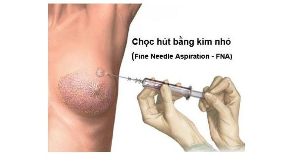
An ultrasound-guided aspiration technique for the diagnosis or treatment of breast cysts.When performing breast cyst aspiration, the doctor will use a small needle to poke the breast cyst and draw out the fluid. If the aspirated fluid does not contain blood, the tumor completely disappears after aspiration, then this is a breast cyst, the patient does not need to do further tests. On the contrary, if the aspirated fluid contains blood and the tumor does not disappear completely, the doctor will send the aspirate for testing, imaging and further treatment.

2.2 Ultrasound-guided breast cyst aspiration procedure
PreparePatient completes necessary documents and procedures; The patient informs the doctor about the drug he is using, the allergy (if any), especially to the anesthetic. Patient informs doctor about recent health situation. Perform
Patient lying supine upright or slightly tilted to one side depending on the doctor's instructions; Selection of the entry route: The doctor uses ultrasound to find the lesion, locate the needle puncture site, and then disinfect the skin of the puncture site. In some cases, the doctor may administer a local anesthetic to relieve the patient's pain. Approach to the lesion: Percutaneous needle is inserted into the center of the mammary cyst under ultrasound guidance. Then, suction out the fluid in the breast cyst. End of intervention: Withdraw the needle, clean the puncture site, apply pressure to prevent bleeding, cover the puncture site with gauze. Finally, the collected specimens will be taken to the pathology laboratory for examination under the microscope.
2.3 Complications and how to deal with them
Bleeding: heavy bleeding (usually rare). To avoid the risk of bleeding, the doctor may apply a tight compression bandage after the procedure, and repeat the ultrasound 1 hour later. Infection: A rare complication. A precaution is to carefully disinfect before performing the technique. At the same time, prophylactic antibiotics can be used if necessary after aspiration. Local anesthetic poisoning: rare due to low dose, if it occurs, the doctor will treat according to the anesthetic poisoning protocol Breast bruising: This condition usually disappears on its own after a few weeks. In case the lesion is too deep close to the chest wall, the aspiration technique can cause pneumothorax or pleural bleeding. For treatment, if suspected, chest X-ray, ultrasound examination and pleural drainage should be placed if indicated; Scars: There is usually little or no scarring on the skin at the puncture site. Scars will fade over time.2.4 Advantages of ultrasound-guided mastectomy
This diagnostic method has many advantages such as:Ultrasound sees the needle position inside the lesion helps the doctor insert the needle into the right lesion, can change the direction of the needle or bring the needle to many different areas of the cyst 🡪 breast allows for more and more accurate specimen collection; Quick diagnosis. Safe, low risk of accidents. High precision. Simple to perform, can repeat the test as needed. Equipment for performing simple techniques. The technique of breast cyst aspiration under ultrasound guidance is quite simple and safe, with few complications. However, the patient still needs to strictly follow all the instructions of the doctor during the procedure in order for the procedure to take place quickly and accurately.

Examination and consultation with an oncologist. Breast cancer screening by bilateral breast ultrasound and mammogram. Officially put into operation on January 7, 2012, Vinmec has become a prestigious address in breast cancer screening with:
Team of highly qualified and experienced doctors. Comprehensive professional cooperation with domestic and international hospitals: Singapore, Japan, USA, etc. Comprehensive treatment and care for patients, multi-specialty coordination towards individualizing each patient. Having a full range of specialized facilities to diagnose the disease and stage it before treatment: Endoscopy, CT scan, PET-CT scan, MRI, histopathological diagnosis, gene-cell testing, .. There are full range of mainstream cancer treatment methods: surgery, radiation therapy, chemotherapy, stem cell transplant...
Please dial HOTLINE for more information or register for an appointment HERE. Download MyVinmec app to make appointments faster and to manage your bookings easily.


