This is an automatically translated article.
The human brain is one of the largest and most important organs of the body. The structure of the human brain is also quite complex. In this article we will provide you with the most basic understanding of the structure of the human brain.1. Overview of the human brain
The brain is considered the jewel and also the most complex part of the body. This 1.36kg organ is the seat of intelligence, the interpreter of the senses, the initiator of body movements and the controller of behavior. Housed in the cortex of bone and washed away by protective fluid, the brain is the source of all the qualities that define us as a person.For centuries, scientists and philosophers have been fascinated by the brain, even considering the brain almost incomprehensible. However, now scientists are gradually approaching its secrets. Scientists have learned more about the brain in the past 10 years than in all previous centuries because of the increasing pace of research in the behavioral and neurosciences and the development of research techniques. new. At the forefront of research into the brain and other elements of the nervous system is the National Institute of Neurological Disorders and Stroke (NINDS), which conducts and supports scientific research in the United States and around the world. gender.
2. Structure of the brain
The brain holds many important functions, controlling all bodily functions, interpreting information from the outside world, and embodying the spirit and soul. Intelligence, creativity, emotions, and memory are just a few of the many things that are governed by the brain.The brain receives information through our five senses: sight, smell, touch, taste and hearing - often multiple senses at once. It gathers messages in a way that makes sense to us and can store that information in our memory. The brain controls our thinking, memory and speech, the movements of our arms and legs, and the function of many organs in our bodies.
The central nervous system (CNS) includes the brain and spinal cord. The peripheral nervous system (PNS) consists of spinal nerves branching from the spinal cord and cranial nerves branching from the brain.
All parts of the brain work together, but each part has its own special properties.

Bộ não người được coi là viên ngọc quý và cũng là bộ phận có cấu tạo phức tạp nhất của cơ thể
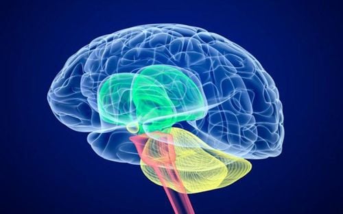
The cerebrum is the largest part of the brain and includes the right and left hemispheres of the brain. It performs higher functions such as tactile, visual, and auditory interpretation, as well as speech, reasoning, emotions, learning, and fine control of movements. Cerebellum: located below the cerebrum. Its function is to coordinate muscle movements, maintain posture and balance. Brainstem: acts as a relay hub connecting the cerebellum and cerebellum to the spinal cord. It performs many automatic functions such as breathing, heart rate, body temperature, wake and sleep cycles, digestion, sneezing, coughing, vomiting and swallowing. 2.2. Right and left brain The brain is divided into two halves: the right and left hemispheres. They are connected by a bundle of fibers called corpuscles that transmit messages from one side to the other. Each hemisphere controls the opposite side of the body. If the stroke occurs in the right side of your brain, your left arm or leg may become weak or paralyzed.
Not all hemisphere functions are shared. In general, the left hemisphere of the brain controls speech, comprehension, arithmetic, and writing. The right hemisphere of the brain controls creativity, spatial ability, art, and musical skills. The left hemisphere of the brain dominates hand use and language in about 92% of people.
In general, the hemisphere of the brain is responsible for language and speech. The right hemisphere of the brain plays a large part in visual information interpretation and spatial processing. In about one-third of left-handed people, speech function may be located on the right side of the brain. Left-handed people may need special testing to determine if the center of their voice is on the left or right side before any surgery in that area is performed.
Aphasia is a language disorder that affects speech production, comprehension, reading or writing, caused by brain injury - most commonly from stroke or trauma. The type of aphasia depends on the area of the brain that is damaged.
Broca's area: located in the left frontal lobe. If this area is damaged, one may have difficulty moving the tongue or facial muscles to produce the sound of speech. The person can still read and understand spoken language but has difficulty speaking and writing (i.e. forming letters and words, not writing in lines) - known as Broca's aphasia. Wernicke's area: located in the left temporal lobe. Damage to this area causes Wernicke's aphasia. Individuals can say long sentences that make no sense, add unnecessary words, and even create new words. They can make speech sounds, however they have difficulty understanding speech and are therefore not aware of their own errors.
Bộ não người gồm đại não, tiểu não và thân não
Frontal lobe: Just below the forehead is the largest lobe of the lobes. It coordinates advanced behaviors such as motor skills, problem solving, judgment, planning, and focusing attention. Parietal lobes: The parietal lobes are located at the upper back of our brain, and control complex behavior, including senses such as vision, touch, body perception, and spatial orientation. It plays an important role in the integration of sensory information from different parts of our body, the knowledge of numbers and their relationships, in the manipulation of objects. Sections related to visual-spatial processing, language comprehension, body construction, positioning and movement, inattention/inattention, left-right discrimination, and perception/ self-knowledge. Occipital lobes: The occipital lobes are located at the back of our brain, and are involved in visual processing, such as visual recognition, visual attention, spatial analysis (moving and movement). in a 3-D world) and visual perception of body language; such as poses, expressions, and gestures. Temporal lobes: The temporal lobes are located near your ears, and are associated with controlling your perception and recognition of auditory stimuli (including the ability to focus on a sound among many other things). different sounds, like listening to a voice in a crowd at a party), verbal comprehension, verbal memory, visual memory, and language production (including fluency and find words), general knowledge, and autobiographical memories. 2.4. The cerebral cortex The surface of the cerebrum is called the cerebral cortex. It has a meandering shape with hills and valleys. The cerebral cortex contains 16 billion neurons (the cerebellum has 70 billion = 86 billion total) arranged into specific layers. The neuronal organelles give the cortex a gray-brown color, hence the name - gray matter (Figure 4). Beneath the cerebral cortex are long nerve fibers (axons) that connect regions of the brain - called white matter.
Folding of the cerebral cortex increases the surface area of the brain allowing more neurons to fit inside the skull and allowing higher functions to be performed. Each fold is called a gyrus, and each groove between the folds is called a fold. There are names for the folds and grooves that help identify specific brain regions.
2.5. Deep structure Pathways called white matter tracts connect regions of the cerebral cortex. Messages can travel from one gyrus to another, from one lobe to another, from one side of the brain to another, and to structures deep in the brain
Hypothalamus: located on the floor of the second ventricle three and is the master controlling organ of the autonomous system. It plays a role in controlling behaviors such as hunger, thirst, sleep, and sexual response. It also regulates body temperature, blood pressure, emotions, and hormone secretion. Pituitary gland: The main role of the pituitary gland is an important link between the nervous system and the endocrine system. The pituitary gland releases many hormones that influence growth, metabolism, sexual and sexual development, promote bone and muscle growth, and response to stress. It is connected to the hypothalamus and is about the size of a pea. It is located in the center of the skull, just behind the bridge of the nose.. Pineal gland: located behind the third ventricle. It helps regulate the body's internal clock and circadian rhythm by secreting melatonin. It has several roles in sexual development. The thalamus: serves as a relay station for almost all incoming and outgoing information to the cerebral cortex. It plays a role in pain sensation, attention, alertness, and memory. Basal ganglia: These nuclei work with the cerebellum to coordinate fine movements, such as fingertip movements. Connected to the hippocampus, it plays a role in emotional memory and contains a large number of opiate receptor sites that are involved in rage, fear, and sexual emotions. Limbic system: is the center of our emotions, learning and memory. Included in this system are the gyrus, hypothalamus, amygdala (emotional response) and hippocampus (memory).
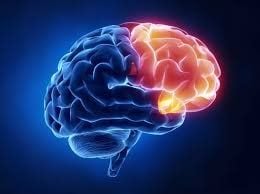
Thùy trán trong bộ não người ngay dưới trán là thùy lớn nhất trong các thùy
A specialized structure in each ventricle, called the choroid plexus, is responsible for the majority of cerebrospinal fluid production. The brain normally maintains a balance between the amount of CSF absorbed and the amount produced. However, disruptions in this system can occur.
There are two ventricles located deep in the cerebral hemispheres called the lateral ventricles. Both connect to the third ventricle through a separate opening known as the foramen of Monro. The third ventricle connects to the fourth ventricle through a long narrow tube called the aqueduct of Sylvius. From the fourth ventricle, CSF flows into the subarachnoid space, where it bathes and cushions the brain. CSF is recycled (or absorbed) by special structures in the upper sagittal sinus called the arachnoid villi.
A balance is maintained between the amount of CSF absorbed and the amount produced. Disruptions or blockages in the system can cause an accumulation of cerebrospinal fluid, which can cause enlargement of the ventricles (hydrocephalus) or cause fluid accumulation in the spinal cord (syringomyelia).
2.7. Skull The purpose of the skull is to protect the brain from injury. The skull is made up of 8 bones fused together by sutures. These bones include the frontal, parietal (2), temporal (2), coccyx, occipital, and ethmoid bones. The face is made up of 14 bone grafts including the maxillary bone, the jawbone, the nose, the roof of the mouth, the lacrimal gland, the lower nose, the mandible and the cane bone.
Inside the skull there are three distinct areas: anterior, middle, and posterior. Doctors sometimes refer to the location of the tumor by these terms, for example meningiomas.
Similar to a cable that goes out to the back of a computer, all the arteries, veins, and nerves exit the base of the skull through holes, called foramina. The large central hole (foramen magnum) is where the spinal cord exits
2.8. Meninges The brain is protected by the skull bones that keep it from being damaged. The skull bones combine with the facial bones to form the skull. Between the skull and the brain is the meninges, which consists of three layers of tissue that protect the brain and spinal cord. From the outermost layer to the inside, the order is: dura mater, arachnoid and membrane.
The sclera: is made up of two layers of white, non-elastic membrane. The outer layer is called the periosteum. One inner layer is the dura mater, which lines the inside of the entire skull and creates small folds or compartments through which parts of the brain are protected and held. Two distinctive folds of the dura mater in the brain are called the cerebellar crescent and the cerebellar tentacle. The cerebellar crescent separates the right and left halves of the brain, and the cerebellar tentacle separates the upper and lower parts of the brain. Arachnoid: The second layer of the meninges is the arachnoid membrane. This membrane is thin and covers the entire brain. There is a space between the dura mater and the arachnoid called the subdural space. The arachnoid is made up of elastic tissue and blood vessels of different sizes. The choroid is the innermost layer close to the surface of the brain and has many blood vessels that go deep into the surface of the brain. The trophoblastic membrane covers the entire surface of the brain, following the folds of the brain. The space that separates the arachnoid membrane from the pia mater is called the subarachnoid space. It is in this area where cerebrospinal fluid circulates in the subarachnoid space.

Màng cứng trong bộ não người được tạo thành từ hai lớp màng màu trắng, không đàn hồi

The vertebral arteries supply the cerebellum, brainstem, and inferior surface of the cerebrum. After passing through the skull, the right and left vertebral arteries join together to form the basilar artery. The basilar and internal carotid arteries “communicate” with each other at the base of the brain known as the Circle of Willis. Communication between the internal carotid system and the vertebral system is an important safety feature of the brain. If one of the major blood vessels is blocked, secondary blood flow can pass through the Circle of Willis and prevent brain damage.
The venous circulation of the brain is very different from the circulation of the rest of the body. Normally arteries and veins run together as they supply and drain water to specific areas of the body. So one would think that there would be a pair of vertebral and internal jugular veins. However, this is not the case with the brain. The main venous collectors are integrated into the sclera to form the venous sinuses - not to be confused with the air sinuses of the face and nose. The venous sinuses collect blood from the brain and deliver it to the internal spherical veins. The superior and inferior ethmoid sinuses drain cerebral blood, and the posterior ethmoid sinuses drain the anterior skull base. All sinuses eventually exit the sigmoid sinus, exiting the skull and forming rectangular veins. These two rectangular veins are essentially the brain's only drainage route.

Tuần hoàn tĩnh mạch của bộ não người rất khác với tuần hoàn của phần còn lại của cơ thể
* Neurons
There are many sizes and shapes of neurons, but they all include a cell body, dendrites, and axons. Neurons transmit information through electrical and chemical signals. Try to visualize the wiring in your home. An electrical circuit is made up of many wires connected in such a way that when a light switch is turned on, the light bulb will light up. An excited neuron transmits its energy to the neurons in its vicinity.
Nerve cells transmit their energy, or “talk,” to each other through a small gap called a synapse. A neuron has many arms called dendrites, which act like antennae to receive messages from other neurons. These messages are passed to the body of the cell, which determines whether the message should be transmitted or not. Important messages are delivered to the end of the axon, where vesicles containing neurotransmitters open into the synapse. Neurotransmitter molecules cross the synapse and bind to special receptors on the receiving neuron, stimulating that cell to transmit the message.
* Glial cells
Are the cells of the brain that provide nerve cells for nourishment, protection, and structural support. There are about 10 to 50 times the number of neurons and are the most common cell type associated with brain tumors.
Astroglia or astrocytes are the caretakers - they regulate the blood-brain barrier, allowing nutrients and molecules to interact with nerve cells. They control homeostasis, protect and repair neurons, scar formation, and also influence electrical impulses. Oligodendroglia cells make a fatty substance called myelin that insulates the axons - allowing electrical messages to travel faster. Spending cells line the ventricles and secrete cerebrospinal fluid (CSF). Microglia are the brain's immune cells, which protect it from invaders and clear debris. They also prune synapses. You should regularly read books, study, do activities that make you use your mind or as simple as playing crossword puzzles... to stimulate your nerve cells and even New brain cells can also be generated.
Please dial HOTLINE for more information or register for an appointment HERE. Download MyVinmec app to make appointments faster and to manage your bookings easily.
References: ninds.nih.gov, nbia.ca, mayfieldclinic.com , healthline.com