This is an automatically translated article.
The article was professionally consulted by Specialist Doctor I Le Hong Lien - Department of Obstetrics and Gynecology - Vinmec Central Park International General Hospital.1. Why prenatal screening?
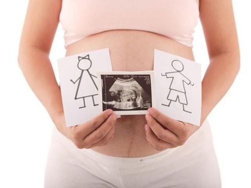
Currently, due to the consumption of harmful chemicals in the diet, the environment, polluted water sources, unhealthy habits... the rate of birth defects increases. Common malformations in infants include:Down syndrome, Edwards syndrome; Slow growth, slow intellectual development; Sexual dysfunction, inability to develop sex; Congenital heart , hypothyroidism ; Cleft lip, cleft palate; Defects in the nervous system, chest, head, chest, face, neck; Deformities of the extremities, malformations of the genitals. Prenatal screening is a series of tests and tests during pregnancy to know the health status of the fetus. The main purpose is to see if the baby is at risk for diseases, birth defects, prenatal screening can be done by many different methods.
Screening and diagnosis when pregnant mothers can detect and intervene early signs to help children develop normally, or reduce consequences for children. In addition, this also helps parents make decisions to keep or abort the fetus if the fetus has too severe defects, is difficult to survive or does not develop well after being born.

2. At what week is prenatal screening?
Prenatal screening is a collection of many tests and checks to monitor the health status and risk of diseases of the baby. Therefore, the implementation of prenatal screening is not mandatory, prenatal screening is completely dependent on the mother's wishes, is usually conducted over many weeks and is not fixed to have antenatal screening at week 2. how many, how much.Prenatal screening has two basic activities: maternal blood test and fetal morphological ultrasound.
In the first 3 months of pregnancy:
Doctors conduct ultrasound of fetal morphology, measure nuchal translucency to detect the risk of Down syndrome. This diagnosis helps doctors detect other problems such as a fetus without a skull, cleft palate, umbilical hernia...; Pregnant women's blood tests determine whether the mother has rubella, HIV and the risk of the fetus having genetic abnormalities; If there are abnormalities, the doctor continues to perform a biopsy of the placenta, to determine the exact genetic status as well as other problems; Pregnant women can also do the Double Test, this is a test done during the 11th to 13th week of pregnancy; If the Double Test results in high-risk results, the pregnant woman will be advised by doctors to perform NIPT test from the 10th week of pregnancy or amniocentesis in the next pregnancy.
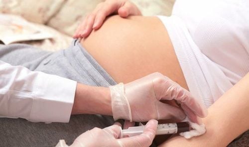
At this time, the fetus is larger, the doctor can look at each part of the baby's body. Screening and diagnosis at this time can detect abnormalities of the baby's nervous system, such as spina bifida, hydrocephalus ...), abnormalities in the cardiovascular system (such as defects in the heart, blood vessels, etc.) , heart valve defects...), abnormalities in the digestive system (such as malformations in the intestines, stomach...), in the genitourinary system, in bones (such as skeletal dysplasia, short limbs...); Blood tests in pregnant women, looking at new infectious diseases in the mother, new chromosomal abnormalities; Triple Test can be done between 15 and 20 weeks of pregnancy; If the test shows a problem, the doctor will do amniocentesis to determine the exact condition of the fetus. However, performing amniocentesis has many risks, so pregnant women from 16 to 24 weeks of age are suitable to conduct it. The implementation of ultrasound, a diagnostic test for fetal malformations, should be done early:
12th to 14th week: Nuchal translucency measurement to predict some chromosomal abnormalities that can cause disease down, diaphragmatic hernia... This is the only time that can make an accurate diagnosis because after 14 weeks, the ultrasound diagnosis is no longer accurate. Week 21 to week 24: Ultrasound helps detect abnormalities in fetal morphology such as cleft lip, cleft palate, organ malformation... Remember, ultrasound only helps detect abnormalities Visibility is completely incapable of diagnosing dysfunction. Weeks 30-32: Ultrasound helps detect some abnormalities in a structural area of the brain, arteries, and heart. At this time, the fetus has grown, there is only one way to give birth to the baby, but understanding the problems of the fetus helps the mother to proactively choose a suitable birth place and set a better care plan for the baby.
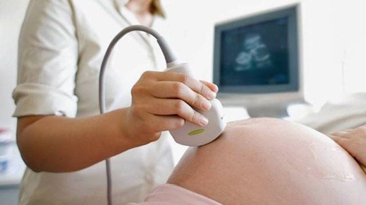
At Vinmec International General Hospital, there is a package maternity service as a solution to help pregnant women feel secure because of the companionship of the medical team throughout the pregnancy. When choosing Maternity Package, pregnant women can:
The pregnancy process is monitored by a team of highly qualified doctors Regular check-ups, early detection of abnormalities The package pregnancy helps to facilitate Convenience for the delivery process Newborns receive comprehensive care, Specialist I Le Hong Lien has been an obstetrician-gynecologist at Vinmec Central Park International Hospital since November 2016. Doctor Lien has over 10 years of experience as a radiologist in the Department of Ultrasound at the leading hospital in the field of obstetrics and gynecology in the South - Tu Du Hospital.
Please dial HOTLINE for more information or register for an appointment HERE. Download MyVinmec app to make appointments faster and to manage your bookings easily.


