This is an automatically translated article.
The article is professionally consulted by Master, Doctor Nguyen Viet Thu - Doctor of Radiology and Nuclear Medicine - Department of Diagnostic Imaging and Nuclear Medicine - Vinmec Times City International General Hospital.High-resolution computed tomography of the lung is a modern imaging method that uses high-intensity X-rays to illuminate the lung parenchyma to capture images of lung structure and perfusion. High-resolution lung CT scan has an important role in supporting the diagnosis and monitoring of treatment response to interstitial lung diseases and bronchial diseases.
1. When should high-resolution computed tomography be indicated?
With the advantage of providing clear image quality, high-resolution computed tomography lung scan, also known as high-resolution lung CT scan, is often preferred to be used in clinical contexts at the hospital. lungs and tracheobronchial tree system. Lesions in lung lobes, bilateral lung lobes, dilated lesions of the air duct system such as alveolar sacs, bronchioles, bronchi, fibrosis and interstitial lung disease have high detection rates on radiographs. Lung CT.High-resolution computed tomography is a relatively safe imaging modality with almost no absolute contraindications. According to many current recommendations, some cases are not recommended to have CT lung or high-resolution lung CT scan, including:
● People with chronic diseases, kidney failure.
● Women who suspect or are pregnant, especially in the first trimester of pregnancy.
● History of drug allergy in general
People with chronic medical conditions that are not well controlled such as bronchial asthma, diabetes.
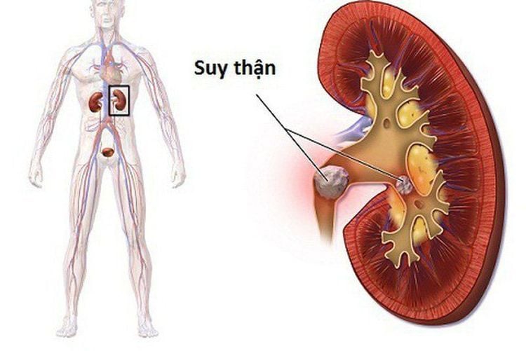
Bệnh nhân suy thận không được khuyến cáo chụp CT phổi
2. High resolution lung computed tomography scan
To ensure patient safety and provide good image quality, high-resolution computed tomography should be performed according to the following steps:● Prepare instruments: CT scan High resolution lung requires a multi-slice computed tomography scanner and an accompanying computer system for image processing. In addition, some medical supplies also need to be prepared including anti-shock medicine boxes, syringes, cotton swabs, physiological saline.
● Prepare the patient: Before entering the high-resolution lung CT scan room, the patient should be consulted and explained clearly and fully the steps to be performed as well as possible complications. When entering the imaging room, the patient should be instructed to change into uniform according to the regulations of each medical facility, remove all jewelry and personal belongings. It is advisable to fast for at least 4 hours before starting a high-resolution lung CT scan. In some hospitals, a family member may be in the room with the patient. In case the patient is too nervous and agitated, sedation should be prescribed.

Bệnh nhân có thể dùng thuốc an thần nếu có biểu hiện lo lắng hay kích động
● Expose the survey area including the entire ribcage to below the diaphragm.
● Perform a lung scan with each slice extending from the top of the lung to the base of the lung, including the costophrenic angle. The thickness and spacing between slices vary depending on the settings of each machine, typically about 1-2 mm for each slice and 10-15 mm between two consecutive slices.
● Do not use contrast medium in this case.
● Change the observation windows in turn, including mediastinal window, lung parenchyma window.
● Conduct evaluation and read the results of the obtained films.
3. Some lesions on high-resolution computed tomography of the lung
High-resolution computed tomography film has outstanding advantages in investigating abnormalities in the lung parenchyma and bronchial system. Several types of lesions can be observed on high-resolution computed tomography (CT) scans of the lungs, including:● Injury in the lobules of the lung: The lobules of the lung are considered the structural and functional units of the mitral lung. Each lung lobe consists of terminal bronchioles and alveolar sacs that are the smallest unit of the air duct system. The lung lobules are separated from each other by a connective tissue layer. Lesions are often observed in the central lobules of the lung, often caused by inflammatory diseases in the lungs such as pneumonia, bronchiolitis, and emphysema. In addition, the lesions observed in the peripheral lymphatics are often suggestive of malignant lesions metastasized from other organs, pulmonary edema.

Viêm phổi là một tổn thương thường được quan sát thấy tại vị trí trung tâm tiểu thùy phổi
Low-density lesions: On high-resolution computed tomography of the lung, low-density tissues are usually air-filled lesions. Therefore, when observing lung parenchyma areas with reduced density, it is necessary to think of diseases that cause air retention in the lungs such as emphysema, bronchial asthma, air bubbles, air cysts, and bronchiectasis. Emphysema can occur in the central lobules or the entire lobules, accompanied by hypodense images that can no longer be observed with healthy lung parenchyma due to complete destruction. The cocoon may present as a focal hypodense organization with thin or thick walls. When the air cocoon has a wall thicker than 4mm, it is called a cave, which is common in infectious diseases.
High resolution computed tomography lung scan is an imaging method that brings high accuracy in diagnosing lung-related diseases, especially dangerous diseases such as pneumonia, cancer. lung. Accordingly, to achieve the most accurate results, you should choose addresses with a quality CT scanning system.
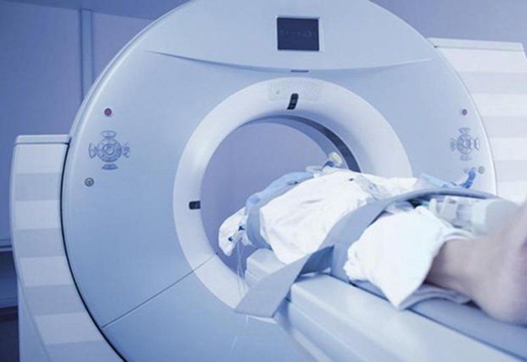
Chụp cắt lớp vi tính phổi có độ phân giải cao là phương pháp chẩn đoán hình ảnh đem lại độ chính xác cao
● Comprehensive patient care and treatment, multi-specialty coordination in the direction of individualizing each patient.
● Having a full range of professional facilities to diagnose the disease and stage it before treatment: Endoscopy, CT scan, PET-CT scan, MRI, histopathological diagnosis, genetic - cytological testing, ...
● There are full range of mainstream cancer treatment methods: surgery, radiation therapy, chemotherapy, stem cell transplant...
Ths.Bs Nguyen Viet Thu has more than 20 years of experience in doing this. Working in the field of diagnostic imaging, former Secretary of the Hanoi Department of Radiology, directing the lower level in the specialty of Diagnostic Imaging. Currently, the doctor is working at the Department of Diagnostic Imaging and Nuclear Medicine - Vinmec Times City International General Hospital.
Currently, all hospitals in Vinmec Health system are applying lung CT scanning technique. Therefore, customers can directly go to Vinmec Health system nationwide to visit or contact the hospital hotline for each area here for support.


