This is an automatically translated article.
The article is professionally consulted by Master, Doctor Le Xuan Thiep - Radiologist - Department of Diagnostic Imaging - Vinmec Ha Long International Hospital. The doctor has extensive experience in the field of diagnostic imaging.Aortic angiography and intervention with digital background erasure is a modern method to help diagnose and treat diseases of the aorta, especially aneurysms and endothelium dissection.
1. What is aortic angiography and intervention?
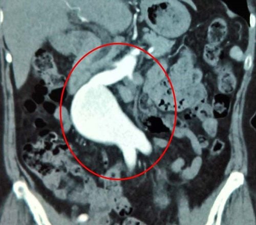
Some pathologies of the aorta are relatively dangerous. Previously, only medical treatment or surgery to replace the abnormal aortic segment had brought negative results. Therefore, at present, aortic angiography and interventional technique is a modern endovascular intervention method with high efficiency and safety for patients.Among the dangerous pathologies in the aorta, the two most common are aneurysms and endothelium dissection. With digitized aortic angiography and intervention, background erasing will use a piece of artificial lumen support with coating (endograft) placed in the lumen of the aorta, covering the aneurysm or the site of dissection. vascular endothelium.
2. When to take aortic scan and intervention?
Asymptomatic thoracic and abdominal aortic aneurysms but aneurysm diameter greater than 55 mm Aortic aneurysms with symptoms of rupture or threat of rupture. Unstable thoracic aortic dissection. Pseudoaneurysm of the thoracic and abdominal aortic aneurysm due to ulcerative plaque threatening to rupture. Perforation of abdominal and thoracic aorta due to trauma Perforation of thoracic and abdominal aorta due to invasive malignancies.3. Contraindications of aortic intervention
Aneurysm, dissection of the ascending aorta Aneurysm, dissection of the aortic arch anterior to the left subclavian artery Diameter of the iliac artery is less than 7 mm End stage renal failure or allergy to contrast iodine. Uncontrolled bleeding disorders. Pregnant women.
Chụp và can thiệp động mạch chủ khi bệnh nhân bị phình động mạch chủ
4. Prepare equipment and drugs before aortic surgery and imaging
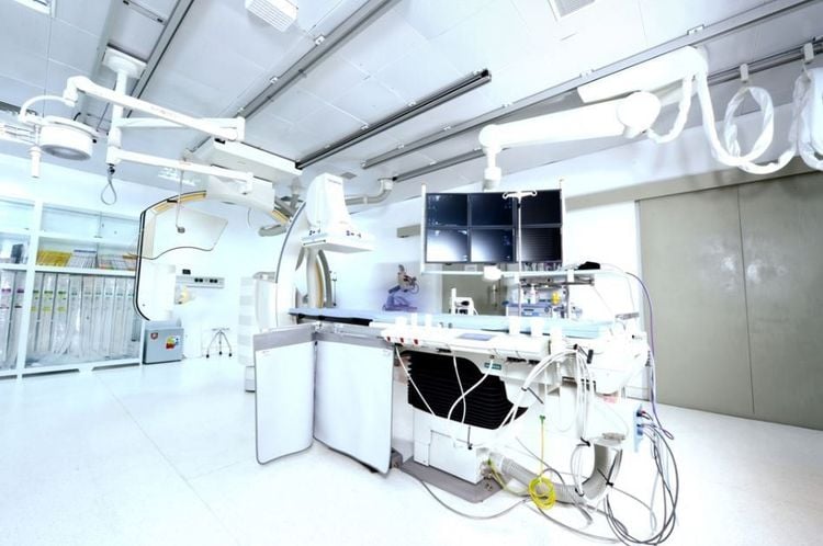
Equipment and vehicles Digital background removal angiography (DSA). Dedicated electric pump Film, film printer, image storage system. Lead shirt, apron to shield X-rays. Medicines to prepare Local anesthetic. Pre-anesthesia and general anesthesia if indicated by the patient. Medicines to treat blood clotting disorders. Water-soluble iodine contrast agent. Antiseptic solution for skin and mucous membranes.5. Prepare the patient before aortic intervention
The patient was thoroughly explained about the procedure and procedure for digitizing aortic angiography and intervention and signed a consent form before the procedure. Patients need to fast, fast for 6 hours before or can drink no more than 50ml of water. After entering the intervention room for aortic imaging and intervention, the patient was lying on his back, and vital signs were monitored such as breathing rate, pulse, blood pressure, electrocardiogram, and arterial blood gases by various machines. . In case the patient is uncooperative or aggressive, a sedative will be prescribed. Disinfect the skin surface with an antiseptic solution and cover with a sterile towel with holes.6. Aortic angiography and interventional procedure digitizing background removal
Anesthesia method The patient is in the supine position and installs an infusion line, usually 0.9% physiological saline solution. In most cases, only local anesthesia is needed. However, with exceptions, it is necessary to inject pre-anesthesia or general anesthesia such as: children under 5 years old, have no sense of cooperation with doctors or patients are too excited, scared... Open the way into the lumen The doctor uses a 21G needle to poke the common femoral artery Insert the catheter into the lumen of the size 5-6F Aortography Bring the catheter system and leads up to the root of the aorta. Replace the catheter with a Pigtail tube. Digital aortic DSA with background erasure to investigate aortic lesions Insert an artificial support system into the aorta Expand the femoral artery with a large 18-22Fr catheter system. Insert the Endograft-coated artificial lumen support over the large 18-22Fr catheter and guided by a stiff wire to the previously identified lesion site. Insertion of Endograft into the lumen of the aorta Determine the position of the Endograft relative to the site of aortic injury through the DSA. Use Endograft fixation system to fix to the lesion site. Use a balloon to shape the lumen inside the Endograft. Then, perform an angiogram to check the circulation after placing the bracket into the lumen of the aorta. Withdraw the entire system from the patient's body. Close the access to the common femoral artery with a closure device. Bandage and immobilize to prevent bleeding.
Máy chụp mạch số hóa xóa nền (DSA)
7. Evaluation of results after angiography and aortic intervention
The procedure of digitizing aortic angiography and intervention with background erasure is considered successful based on the following criteria:Endograft covers the entire damaged aortic lumen and does not obstruct the blood vessels separated from the artery motherboard. There is no interstitial blood flow between Endograft and aortic wall. The large arteries that separate from the aorta have good circulation.
8. Complications and treatment direction after aortic intervention
Infections and Hematomas in the inguinal region Since the aorta's access route needs to be widened compared to other procedures, the risk of infection and hematoma is also higher. The common treatment is medical treatment and monitoring, local care.Injury to the common femoral artery The system of catheters and racks inserted into the femoral artery has a large size of 18-22Fr, increasing the risk of dissection and rupture of the femoral artery. Treat with stenting or surgery. To limit this complication, it is necessary to accurately assess the diameter of the femoral iliac artery before aortic intervention.
Infection of the vascular support frame This is a rare complication, the infection rate is about 0.5 - 1%. Most only need internal treatment, but a few cases need surgery, making an artificial bridge.
Vinmec International General Hospital is a high-quality medical facility in Vietnam with a team of highly qualified medical professionals, well-trained, domestic and foreign, and experienced.
A system of modern and advanced medical equipment, possessing many of the best machines in the world, helping to detect many difficult and dangerous diseases in a short time, supporting the diagnosis and treatment of doctors the most effective. The hospital space is designed according to 5-star hotel standards, giving patients comfort, friendliness and peace of mind.
Please dial HOTLINE for more information or register for an appointment HERE. Download MyVinmec app to make appointments faster and to manage your bookings easily.


