This is an automatically translated article.
The article is professionally consulted by Master, Doctor Ton Nu Tra My - Department of Diagnostic Imaging - Vinmec Central Park International General Hospital.Ultrasound is a modern imaging method used in the examination, diagnosis and treatment of diseases at medical facilities and hospitals. Therefore, now there are many types of ultrasound machines born to meet that need. Besides the advantages of ultrasound, there are also certain limitations of ultrasound.
1. Advantages of ultrasound machine in diagnostic imaging
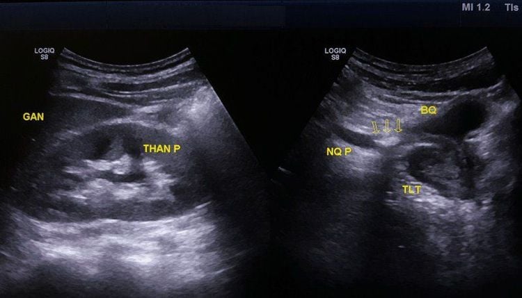
Ultrasound is a non-invasive imaging method that helps doctors diagnose and treat diseases. Conventional ultrasound produces images of thin, flat slices of the body. Advances in modern ultrasound technology today include 3D ultrasound (3D ultrasound) which is able to reconstruct data obtained from sound waves into 3D images. 4D ultrasound (4D ultrasound) is 3D ultrasound that records movement. Accordingly, some advantages of ultrasound machines can be mentioned such as:Helps doctors perform examination of diseases such as: tumors, inflammation, deformities, ... at many locations on the body such as foci of the body. abdomen, pelvis, liver, bile, kidney, breast, neck, uterus, fetus Ultrasound can assess fetal development, especially with 3D, 4D ultrasound, doctors can evaluate early fetal morphological abnormalities. Ultrasound assesses the size and location of stones such as kidney stones, bladder stones, urethral stones...

2. Limitations of ultrasound machines
Besides the advantages of ultrasound, this imaging method also has certain limitations. Specifically:Ultrasound cannot accurately diagnose the intestines and parts that are obscured by the intestines because the sound waves are obstructed by air and vapors. At this time, methods such as CT scan and MRI can be considered by doctors. Ultrasound cannot travel through air. Therefore, organs such as stomach, pancreas, and aorta often need to be combined with other imaging methods for diagnosis. With ultrasound, the doctor can only see the outer surface of the bone, not the inside of the bone. In addition, ultrasound also limits the survey in overweight and obese people,

3. Steps to perform ultrasound
In order for the ultrasound to give the most accurate diagnosis results, the patient should strictly follow the instructions of the sonographer. Accordingly, the ultrasound steps are performed with the following procedure:Step 1. Prepare the patient
The patient should wear comfortable, loose-fitting clothes when going for the ultrasound. In some cases, the doctor may instruct the patient to remove clothing and jewelry in body positions that need to be examined. Some procedures may require the patient to wear a gown to facilitate the examination.
In addition, other preparations will depend on what ultrasound the patient will be having, for example, there are types of ultrasound that require the patient to fast for 6-8 hours before, or some types of ultrasound can Ask the patient to drink several glasses of water within 2 hours, and at the same time hold back from urinating so that the bladder fills with water during examination.
Step 2. How to perform an ultrasound
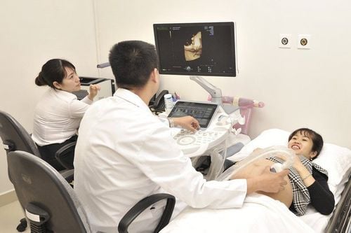
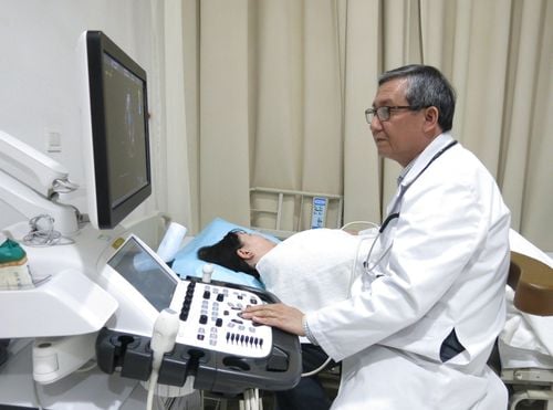
During most ultrasound exams, the patient will be asked to lie on their back on an examination table. The doctor will use a clear, sterile gel and apply it to the body, limiting air between the transducer and the patient's skin. The sonographer will then press the transducer against the patient's skin and scan backward and forward over the areas of the body being examined.
Usually, the sonographer can diagnose the disease based on the ultrasound images right in the time of the procedure and the patient can receive the results immediately after.

Transesophageal echocardiography : A probe is inserted into the esophagus to take pictures of the heart. Transrectal ultrasound: a transducer will be inserted into the rectum of a male patient to view the prostate gland. Transvaginal ultrasound: The transducer will be inserted into the female patient's vagina to view the uterus and ovaries. This procedure is performed only with the consent of the patient. Married or sexually active patients Most ultrasounds are done between 10 minutes and 30 minutes.
Ultrasound is one of the effective imaging methods to help doctors diagnose, determine the stage of the disease to have an effective treatment regimen. In addition, ultrasound also brings many other applications such as therapeutic ultrasound or pregnancy ultrasound to monitor fetal development. Besides a number of advantages, ultrasound also has certain limitations. Therefore, choosing a reputable medical facility with a system of modern medical equipment is very necessary.

Please dial HOTLINE for more information or register for an appointment HERE. Download MyVinmec app to make appointments faster and to manage your bookings easily.


